KUMEL Repository
1. Journal Papers (연구논문)
1. School of Medicine (의과대학)
Dept. of Internal Medicine (내과학)
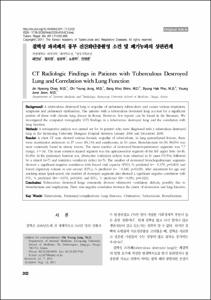
결핵성 파괴폐의 흉부 전산화단층촬영 소견 및 폐기능과의 상관관계
- Alternative Author(s)
- Jung, Chi Young; Jeon, Young June; Rho, Byung Hak
- Journal Title
- Tuberculosis and Respiratory Diseases
- ISSN
- 1738-3536
- Issued Date
- 2011
- Abstract
- Background
A tuberculous destroyed lung is sequelae of pulmonary tuberculosis and causes various respiratory symptoms and pulmonary dysfunction. The patients with a tuberculous destroyed lung account for a significant portion of those with chronic lung disease in Korea. However, few reports can be found in the literature. We investigated the computed tomography (CT) findings in a tuberculous destroyed lung and the correlation with lung function.
Methods
A retrospective analysis was carried out for 44 patients who were diagnosed with a tuberculous destroyed lung at the Keimyung University Dongsan Hospital between January 2004 and December 2009.
Results
A chest CT scan showed various thoracic sequelae of tuberculosis. In lung parenchymal lesions, there were cicatrization atelectasis in 37 cases (84.1%) and emphysema in 13 cases. Bronchiectasis (n=39, 88.6%) was most commonly found in airway lesions. The mean number of destroyed bronchopulmonary segments was 7.7 (range, 4~14). The most common injured segment was the apicoposterior segment of the left upper lobe (n=36, 81.8%). In the pulmonary function test, obstructive ventilatory defects were observed in 31 cases (70.5%), followed by a mixed (n=7) and restrictive ventilatory defect (n=5). The number of destroyed bronchopulmonary segments showed a significant negative correlation with forced vital capacity (FVC), % predicted (r=-0.379, p=0.001) and forced expiratory volume in one second (FEV1), % predicted (r=-0.349, p=0.020). After adjustment for age and smoking status (pack-years), the number of destroyed segments also showed a significant negative correlation with FVC, % predicted (B=-0.070, p=0.014) and FEV1, % predicted (B=-0.050, p=0.022).
Conclusion
Tuberculous destroyed lungs commonly showed obstructive ventilatory defects, possibly due to bronchiectasis and emphysema. There was negative correlation between the extent of destruction and lung function.
Keywords: Tuberculosis, Pulmonary/complications; Lung Diseases, Obstructive; Tuberculosis; Bronchiectasis
- Alternative Title
- CT Radiologic Findings in Patients with Tuberculous Destroyed Lung and Correlation with Lung Function
- Publisher
- School of Medicine
- Citation
- 채진녕 et al. (2011). 결핵성 파괴폐의 흉부 전산화단층촬영 소견 및 폐기능과의 상관관계. Tuberculosis and Respiratory Diseases, 71(3), 202–209. doi: 10.4046/trd.2011.71.3.202
- Type
- Article
- ISSN
- 1738-3536
- Appears in Collections:
- 1. School of Medicine (의과대학) > Dept. of Internal Medicine (내과학)
1. School of Medicine (의과대학) > Dept. of Radiology (영상의학)
- 파일 목록
-
-
Download
 oak-aaa-5109.pdf
기타 데이터 / 858.95 kB / Adobe PDF
oak-aaa-5109.pdf
기타 데이터 / 858.95 kB / Adobe PDF
-
Items in Repository are protected by copyright, with all rights reserved, unless otherwise indicated.