KUMEL Repository
1. Journal Papers (연구논문)
1. School of Medicine (의과대학)
Dept. of Internal Medicine (내과학)
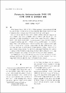
Puromycin Aminonucleoside 투여로 인한 사구체 기저막 및 상피세포의 변화
- Keimyung Author(s)
- Kim, Hyun Chul; Park, Kwan Kyu
- Journal Title
- 대한신장학회지
- Issued Date
- 1998
- Volume
- 17
- Issue
- 6
- Abstract
- Puromycin aminonucleoside(PAN) nephropathy was induced in a group of Sprague-Dawley rat by a single dose of intraperitoneal injection to study an ultrastructural alteration of glomerular anionic sites by stain with poJyethyleneimine(PEI) as a cationic probe and to examine whether proliferation of podocytes occur by immunohistochemical stain for proliferating cell nuclear antigen(PCNA).
The experimental rats developed proteinuria three days after PAN injection. Electron microscopic studies of glomeruli showed the loss of epithelial foot processes, formation of cytoplasmic vacuoles, microvillous formation and increased numbers of lyso-somes in the cytoplasm of podocytes. PEI method seems to selectively stain heparan sulfate proteoglycan in basement membrane and has been widely used to evaluate the changes of basement membrane in human disease as well as in experimental work.
The anionic sites on the basement membrane with foot process fusion were mostly indistinguishable from those seen in control rats, but focal areas of loss or disarray of anionic sites were noted. The anionic sites were not seen on the basement membrane where the overlying epithelium was detached.
It is strongly suggested that proteinuria in FAN nephrosis may be primarily due to a glomerular epithelial lesion, leading Co focal disarray of anionic sites or focai defects in the epithelial covering of the basement membrane. The loss of anionic sites in the basement membrane may be resulted partially from the foot process fusion and mostly from the epithelial detachment. The increased numbers of PCNA positive cells after the injection of PAN is suggestive of possibility of podocytic proliferation or regeneration.
저자는 Sprague-Dawley 흰쥐 35 마리를 대상으로 puromycin aminonucIeoside(PAN)를 복강 내에 주사하여 신 손상을 일으킨 후 사구체 상피세포의 변화, 기저막의 음이온부위의 변화 와 단백뇨와의 관계 및 족세포의 재생여부를 관찰하여 다음과 같은 성적을 얻었다.
PAN 투여 3일 및 7일째 부터 1일 요 단백이 증가하기 시작하여 10일째 293±44mg으로 최 대로 증가하였다. PAN 투여 후 초미형태학적 변화로는 족양돌기의 융합, 리소솜의 증가, 미세 융모성 변화 등이 3일째 부터 관찰되었으며 이러한 소견들은 16일째 부터 회복되기 시작하여 20 일째에는 대체로 정상과 비슷하게 되었다. Po]yethyleneimine을 이용한 음이온부위의 관찰을 통 해 실험군에서 족세포의 융합 자체는 기저막내의 음이온을 소실시키지 않았다. 그러나 족세포가 융합된 부위의 기저막 일부에서 음이온이 불규칙하게 존재하는 경우가 있었으며 족세포가 탈락 된 곳의 기저막에는 음이온이 소실되었다. 융합된 족세포가 복구되면 기저막의 음이온의 구조도 정상 상태로 회복되었다. 상피세포의 표면에도 음이온이 존재하며 그 변화는 기저막의 음이온 변 화와 유사하였다. PCNA 양성세포 수는 대조군에서 1.06개, PAN 투여군에서는 투여 7일째 평 균 3.09개로 유의하게 증가하였다(P<0.05). PCNA 양성세포는 대부분 상피세포였고 그 수는 PAN 투여후 증가하였으며 그것은 상피세포의 증식이 있음을 시사하는 소견의 하나로 생각되었다.
다만 형태학적으로는 음이온 부위가 회복되었으나 단백뇨는 어느정도 지속되었는데 과연 전자 현미경으로 관찰되지 않는 틈이 있는지 혹은 형태학적으로는 회복된 전하장벽이 기능적으로는 역할을 충분히 못하는지의 여부 등은 추후 더 연구되어야 할 것으로 생각된다.
- Alternative Title
- Morphological Changes in Glomerular Epithelial Cells and Basement Membranes in Puromycin Aminonucleoside Induced Nephropathy
- Publisher
- School of Medicine
- Citation
- 최찬오 et al. (1998). Puromycin Aminonucleoside 투여로 인한 사구체 기저막 및 상피세포의 변화. 대한신장학회지, 17(6), 853–865.
- Type
- Article
- ISSN
- 1225-0015
- Appears in Collections:
- 1. School of Medicine (의과대학) > Dept. of Internal Medicine (내과학)
1. School of Medicine (의과대학) > Dept. of Pathology (병리학)
- 파일 목록
-
-
Download
 oak-bbb-02387.pdf
기타 데이터 / 1.95 MB / Adobe PDF
oak-bbb-02387.pdf
기타 데이터 / 1.95 MB / Adobe PDF
-
Items in Repository are protected by copyright, with all rights reserved, unless otherwise indicated.