KUMEL Repository
1. Journal Papers (연구논문)
1. School of Medicine (의과대학)
Dept. of Internal Medicine (내과학)
초산납을 투여한 흰쥐 신장의 초미형태학적 변화
- Keimyung Author(s)
- Kim, Hyun Chul; Park, Kwan Kyu
- Department
- Dept. of Internal Medicine (내과학)
Dept. of Pathology (병리학)
Institute for Medical Science (의과학연구소)
- Journal Title
- 대한병리학회지
- Issued Date
- 1996
- Volume
- 30
- Issue
- 2
- Abstract
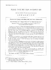
- This study was carried out to investigate the ultrastructural findings of rats after administration of 0.5% lead acetate with drinking water. The Sprague-Dawley rats were divided into control and experimental groups. The control group was composed of 12 rats and was orally administered with 0.5% sodium acetate. The experimental group was composed of 36 rats and orally administered with 0.5% lead acetate. Two rats in the control group and four rats in the experimental group were sacrificed on day 2, and week 1, 2, 4, 6 and 8 after administration. The kidney was extirpated and examined by electron microscopy. The results obtained were as follows: The blood lead concentration in the experimental group began to increase from the second day after administration and it increased gradually until the 6th week and it decreased at the 8 week. The urinary excretion of delta-ALA also increased from the secondary and gradually increased up to the 8th week. On electron microscopic examination, the proximal tubular cells showed fat droplets, dilatation of the endoplasmic reticulum, mitochondrial swelling, increased numbers of secondary lysosomes and myelin figure-like residual bodies and intranuclear inclusion bodies. All these findings peaked at the eighth week after administration. Ultrastructural findings after Timm sulphide silver reaction revealed the lead granules in the proximal tubular lumen and between the microvilli of the proximal tubular cells without membrane-bounded. It can be concluded that most of the changes of micro-organelles are compatible with degenerative changes of lead exposure and passive diffusion of lead granules are involved in the proximal tubular cells.
Key Words: Lead; Kidney; Timm sulphide; Electron microscopy
저자는 Sprague-Dawley 흰주 48마리를 대상으로 증류수에 초산납을 섞어 0.5%의 농도로 마시게한 후 신장을 적출하여 그 변화를 초미형태적으로 관찰하고 Timm 반응을 시킨 후 납의 축적위치를 투과전자현미경으로 관찰한 성적을 요약하며 다음과 같다.
1. 혈중 납 농도는 대조군에서는 0.92㎍/ml 이었고 초산납 투여 후 2주째군에서 평균 3.29㎍/ml로 최고치를 나타내었다.
2. 뇨중 δ-aminolevulinic acid 대조군은 9.8mg/creatinine mg이었고 2일군부터 증가되기 시작하여 8주군에서 71.7mg/creatinine을 나타내었다.
3. 전자현미경적 소견으로는 주로 근위세뇨관에서 많은 변화가 이루어 졌으며 지방적의 증가, 내형질 세망의 확장 및 공포화, 미토콘드리아의 종창, 리소솜 및 수초모양 잔류체의 증가와 핵내봉입체의 출현등이며 이러한 변화들은 초산납 투여후 8주째군에서 가장 저명하였다.
4. Timm 반응을 일으킨 후의 투과전자현미경적 소견은 납 과립들이 주로 근위세뇨관강에 존재해있었고 일부는 원형질막에 둘러싸이지 않은채 근위세뇨관세포의 미세융모 사이에 존재해 있어서 단순 확산에 의한 재흡수로 사료되었다.
이상의 소견을 종합해 보면 흰쥐에게 초산납을 경구 투여했을 때의 주된 손상부위는 근위세뇨관으로 생각되고, 초기에는 가역적인 변화이나 나중에는 핵의 면화 및 파괴까지 초래되는 비가역적인 변화까지 초래되었다. 납의 축적 부위는 사구체를 통해 뇨강으로 빠져 나간뒤 근위세뇨관세포에서 단순확산에 의한 재흡수과정을 통해 근위세뇨관 세포내로 함입된다고 믿어지나 세포내 정확한 소기관의 축적부위에 관해서는 추후 더 연구되어야 할 것으로 사료된다.
- Alternative Title
- Ultrastructural Changes in Rat Kidney after Lead Acetate Administration.
- Publisher
- School of Medicine
- Citation
- 김현철 et al. (1996). 초산납을 투여한 흰쥐 신장의 초미형태학적 변화. 대한병리학회지, 30(2), 73–88.
- Type
- Article
- ISSN
- 0379-1149
- 파일 목록
-
-
Download
 oak-bbb-1169.pdf
기타 데이터 / 32.02 MB / Adobe PDF
-
Items in Repository are protected by copyright, with all rights reserved, unless otherwise indicated.