KUMEL Repository
1. Journal Papers (연구논문)
1. School of Medicine (의과대학)
Dept. of Internal Medicine (내과학)
내시경 점막 절제술 및 내시경 점막하 박리술 시행 중 발생한 위장천공의 비수술적 치료
- Keimyung Author(s)
- Cho, Kwang Bum; Jang, Byoung Kuk; Chung, Woo Jin; Park, Kyung Sik; Hwang, Jae Seok
- Department
- Dept. of Internal Medicine (내과학)
- Journal Title
- 대한소화기내시경학회지
- Issued Date
- 2008
- Volume
- 37
- Issue
- 2
- Abstract
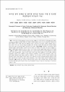
- Background/Aims: Endoscopic Mucosal Resection (EMR) and Endoscopic Submucosal Dissection (ESD) are novel techniques used for the treatment of early gastric cancer and precancerous lesions of the stomach. However, complications such as bleeding and perforation may occur during the procedure, and these complications may raise the morbidity and mortality rates. EMR/ESD-induced perforations can be treated with conservative medical or non-surgical methods. Furthermore, an increasing number of reports have addressed conservative management of EMR/ESD-induced perforations. We evaluated the effectiveness and safety of implementing conservative treatment for perforations associated with EMR and ESD. Methods: We reviewed 482 patients with 507 lesions who underwent EMR or ESD due to early gastric cancers or gastric adenomas between February 2003 and December 2007. We identified 14 perforations occurring as complications of EMR/ESD and investigated their clinical outcomes. Results: Fourteen perforations (14/507 [2.8%]) occurred, 11 of which were immediately clipped during the procedure, and 3 of which were diagnosed after the procedure when free air was visualized on the radiograph. All patients were managed conservatively with fluid resuscitation and antibiotics (mean, 5.8 days). They recovered without surgery and were discharged in stable condition at a mean of 7.2 days post-procedure. Conclusions: Endoscopic clip application might be an effective and safe option for conservative management of EMR/ESD-induced perforations.
목적: 내시경 점막 절제술(endoscopic mucosal resection, EMR) 및 내시경 점막하 박리술(endoscopic submucosal dissection, ESD)은 점막에 국한된 조기 위암 및 전암성 병변에 대하여 비교적 비침습적인 치료법으로서 현재 시행되고 있으나 출혈, 천공 등의 합병증이 생길 수 있으며 이 중 천공은 사망률, 이환율을 높일 수 있다. 따라서 저자 들은 시술 중 천공이 발생했을 경우 내시경을 이용한 클립 봉합 등의 비수술적 방법을 통한 치료의 효과 및 안전성에 대해서 평가해 보고자 하였다. 대상 및 방법: 계명대학교 동산의료원 소화기내과에서 2003년 2월부터 2007년 12월까지 점막을 침범한 조기 위암 및 위 선종을 가진 환자 중 EMR 또는 ESD를 시행한 482명, 507병소를 대상으로 하였고, 이 중 천공이 발생한 14예에 대해서 후향적으로 분석하였다. 결과: 총 14예에서 천공이 발생하였다(14/507, 2.8%). 천공이 발생한 환자들의 연령은 68세였으며, 병변의 크기는 평균 35.6 mm이었다. 천공이 발생한 위치는 분문부 1예, 전정부 4예였으며, 나머지 9예는 체부에서 발생하였다. 천공의 크기는 12예에서 5 mm 이하였으나, 2예에서는 크기가15 mm 이상이었다. 11예는 시술 중에 천공을 발견하여 클립 봉합을 하였으나, 3예에서는 시술 후 방사선 검사에서 천공이 발견되어 클립 봉합 없이 보존적 치료만 하였다. 보존적 치료로 금식, 정맥 내 수액 공급 및 항생제 치료(평균 5.8일)를 실시하였다. 14예 모두에서 수술적 치료 없이 증상이 호전되었고, 시술 후 평균 7.2일간 재원 후 특별한 증상 없이 퇴원하였다. 결론: 내시경 점막 절제술 및 내시경 점막하 박리술 시행 중 발생한 천공은 내시경을 이용한 즉각적인 클립 봉합 등의 비수술적 치료로 비교적 안전하게 치료될 수 있으므로 천공 발생 시 우선적으로 시행할 수 있다.
- Alternative Title
- Nonsurgical Treatment of Gastric Perforation Complicated by Endoscopic Mucosal Resection and Endoscopic Submucosal Dissection
- Publisher
- School of Medicine
- Citation
- 이석근 et al. (2008). 내시경 점막 절제술 및 내시경 점막하 박리술 시행 중 발생한 위장천공의 비수술적 치료. 대한소화기내시경학회지, 37(2), 97–104.
- Type
- Article
- ISSN
- 1225-7001
- Appears in Collections:
- 1. School of Medicine (의과대학) > Dept. of Internal Medicine (내과학)
- 파일 목록
-
-
Download
 oak-bbb-2228.pdf
기타 데이터 / 2.09 MB / Adobe PDF
-
Items in Repository are protected by copyright, with all rights reserved, unless otherwise indicated.