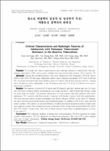
청소년 폐결핵의 임상적 및 영상학적 특징: 재활동성 결핵과의 관련성
- Alternative Author(s)
- Kim, Yeo Hyang; Jung, Chi Young; Lee, Hee Jung
- Journal Title
- 소아알레르기 및 호흡기학회지
- ISSN
- 1225-679x
- Issued Date
- 2012
- Abstract
- Purpose: To evaluate the clinical characteristics and radiologic patterns of adolescents with pulmonary tuberculosis (TB), and to assess whether they are related with primary TB or reactive TB.
Methods: Among the enrolled patients who were diagnosed with pulmonary TB from March 2000 to May 2011, 36 with plain radiography and/or chest computed tomography (CT) were reviewed. We reviewed retrospectively their medical charts to collect clinical data and past history. Among these 36 patients, plain radiography of the 36 patients and chest CT of the 34 patients were retrospectively evaluated.
Results: The patients consisted of 18 males and 18 females, and their median age was 14 years old. The most common clinical presentation was cough and fever. Half of them had chronic cough for more than two weeks. Ten patients had history of close contact with adult patients with active pulmonary TB: 7 patients with their parents, 2 patients with friends, 1 patient with their grandmother. The most frequent pattern of plain radiography was pleural effusion (16/36). In the chest CT findings, all cases showed parenchymal lesions and lymphadenopathy. In addition, 91% of the cases showed acinar nodules. The pattern of pleural effusion revealed associated ipsilateral pleural lymph node and subpleural nodule. Rim enhancement and calcification of the lymph node demonstrated 9% (3/34) and 12% (4/34), respectively. Only two of them showed typical hilar lymphadenopathy in chest X ray and CT.
Conclusion: The radiologic findings of adolescents with pulmonary TB show patterns for rather reactive than primary TB. For diagnosis of adolescent pulmonary TB, chest CT is more helpful than that of plain radiography.
목적: 이 연구에서는 청소년 폐결핵 환자들의 임상적 및 영상학적 특징을 조사하고, 이를 바탕으로 청소년 폐결핵이 초감염 결핵과 재활동성 결핵 중 어떤 형태로 많이 발생하는지 알아 보고자 하였다.
방법: 36명의 환자를 대상으로 의무 기록과 영상학적 검사 결과를 후향적으로 검토하였고, 36명의 환자에서 흉부 X선 영상을, 34명의 환자에서 흉부 전산화 단층촬영 영상을 확인할 수 있었다.
결과: 대상 환자군의 나이 중간값은 14(10-17세)였고, 성별은 남자 18명, 여자 18명이었다. 가장 흔한 임상 양상은 기침과 발열이었고 21명 (50%)에서 2주 이상의 기침이 있었다. 활동성 결핵 환자와 접촉력이 있었던 환자는 10명(25%)으로, 7명은 부모가 결핵 환자였고, 2명은 친구, 1명은 할머니가 결핵 환자였다. 흉부 X선에서 가장 많은 빈도를 보인 양상은 흉수로 16명(44%)에서 보였고, 폐실질 병변 없이 정상 흉부 X선 소견을 보인 경우가 10명(27%)이었다. 흉부 전산화 단층촬영 영상에서는 모든 환자가 폐실질 병변과 림프절병증이 관찰되었다. 폐실질의 주병변은 폐상엽이었고, 폐실질 병변 중 가장 빈도가 많은 것은 꽈리 결절이었으며, 31명(91%)에서 보였다. 그 외 경화, 육아종, 기관지 확장증, 젖빛유리혼탁, 섬유화 등이 관찰되었다. 림프절병증이 가장 많이 보인 곳은 우상부 기관옆 림프절이었고, 30명(88%)에서 두 곳 이상의 림프절병증이 있었다. 2단지명 (6%)에서만 흉부 X선 사진과 흉부 전산화 단층촬영 영상에서 전형적인 폐문부 림프절 비대 소견을 보였다.
결론: 국내 청소년 폐결핵은 영상학적으로 초감염보다는 재활동성 감염의 패턴을 보이며, 청소년 폐결핵의 진단에는 흉부 X선 검사보다 흉부 전산화 단층촬영이 더 도움이 된다.
- Alternative Title
- Clinical Characteristics and Radiologic Patterns of Adelescents with Pulmonary Tuberculosis: Relevance to the Reactive Tuberculosis
- Publisher
- School of Medicine
- Citation
- 강석진 et al. (2012). 청소년 폐결핵의 임상적 및 영상학적 특징: 재활동성 결핵과의 관련성. 소아알레르기 및 호흡기학회지, 22(2), 163–170. doi: 10.7581/pard.2012.22.2.163
- Type
- Article
- ISSN
- 1225-679x
- 파일 목록
-
-
Download
 oak-bbb-2307.pdf
기타 데이터 / 554.78 kB / Adobe PDF
-
Items in Repository are protected by copyright, with all rights reserved, unless otherwise indicated.