KUMEL Repository
1. Journal Papers (연구논문)
1. School of Medicine (의과대학)
Dept. of Orthopedic Surgery (정형외과학)
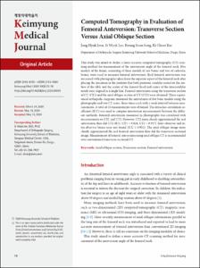
Computed Tomography in Evaluation of Femoral Anteversion: Transverse Section Versus Axial Oblique Section
- Keimyung Author(s)
- Jeon, Jong Hyuk; Lee, Si Wook; Song, Kwang Soon; Bae, Ki Cheor
- Department
- Dept. of Orthopedic Surgery (정형외과학)
- Journal Title
- Keimyung Medical Journal
- Issued Date
- 2020
- Volume
- 39
- Issue
- 2
- Abstract
- This study was aimed to define a more accurate computed tomography (CT) scanning method for measurement of the anteversion angle of the femoral neck. Five models of the femur, consisting of three models of saw bones and two of cadaveric bones, were used to measure femoral anteversion. Real femoral anteversion was measured with photographs taken from the superior aspect of the femoral neck after placing the specimen in the position that both posterior condyles rested on the surface of the table and the center of the femoral head and center of the intercondylar notch were aligned in a single line. Femoral anteversion using the transverse section of CT (CT1) and the axial oblique section of CT (CT2) were obtained. Three experienced orthopedic surgeons measured the anteversion of five bone models using the photographs and two CT scans, three times each with a week interval between measurements. A total of 45 measurements were obtained. The intraclass correlation coefficient (ICC) was used to compare anteversion measurements between the different methods. Femoral anteversion measured in photographs was correlated with measurements on CT1 and CT2. However, CT2 more closely approximated the real anteversion than did CT1 (ICC; CT1 = 0.824, CT2 = 0.937). Inter-observer and intra-observer biases were not found (ICC ≥ 0.952). The axial oblique image more closely approximated the real femoral anteversion than did the transverse sectional image. Measurement of femoral anteversion using axial oblique CT is recommended over conventional transverse sectional CT.
- Citation
- Jong Hyuk Jeon. (2020). Computed Tomography in Evaluation of Femoral Anteversion: Transverse Section Versus Axial Oblique Section. Keimyung Medical Journal, 39(2), 72-78. doi: 10.46308/kmj.2020.00045
- Type
- Jounral
- Source
- https://www.e-kmj.org/journal/view.php?doi=10.46308/kmj.2020.00045
- Appears in Collections:
- 2. Keimyung Medical Journal (계명의대 학술지) > 2020
1. School of Medicine (의과대학) > Dept. of Orthopedic Surgery (정형외과학)
- 파일 목록
-
-
Download
 KMJ-2020-203.pdf
기타 데이터 / 2.56 MB / Adobe PDF
KMJ-2020-203.pdf
기타 데이터 / 2.56 MB / Adobe PDF
-
Items in Repository are protected by copyright, with all rights reserved, unless otherwise indicated.