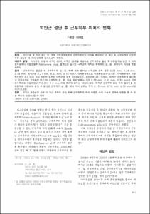
외안근 절단 후 근부착부 위치의 변화
- Affiliated Author(s)
- 이세엽
- Alternative Author(s)
- Lee, Se Youp
- Journal Title
- 대한안과학회지
- ISSN
- 0378-6471
- Issued Date
- 2006
- Abstract
- Purpose: This study examines the change in distance from the corneal limbus to the insertion of the rectus muscles before and after disinsertion and retraction with a pair of fixation forceps during strabismus surgery.
Methods: In 38 strabismus patients, on 30 medial rectus muscles and 38 lateral rectus muscles, before and after disinsertion and retraction with a pair of fixation forceps, the distances from the corneal limbus to the upper, middle and lower parts of the insertion of the medial and lateral rectus muscles marked with methylene blue solution were measured.
Results: The distances between the corneal limbus and upper, middle and lower parts of the insertion after the disinsertion were reduced on, average 0.23 mm, 0.28 mm, and 0.18 mm, for the medial rectus muscle, respectively, and 0.21 mm, 0.28 mm, and 0.15 mm, for the lateral rectus muscle, respectively (P<0.05). The percentage of cases in which the advance from the corneal limbus to the insertion was greater than 0.5 mm was 33.3% for the medial rectus muscle, and 21.1% for the lateral rectus muscle. The lateral rectus muscle was disinserted, fixed with a pair of forceps, and subsequently, the distance from the upper, middle and lower parts to the corneal limbus were reduced to 0.36 mm, 0.43 mm, and 0.30 mm, respectively (P<0.05). The percentage of cases that advanced more than 1.0 mm was 13.2 %. The changes in distance from the upper, middle, and lower parts of insertion toward the limbus after disinsertion and retraction were 0.15 mm 0.15 mm, 0.16 mm, respectively (P<0.05).
Conclusions: When performing the recession of the lateral rectus muscle, disinsertion of the rectus muscle, may result in a change of the site of insertion, which in turn might influence the outcome of strabismus surgery.
목적 : 사시수술 중 직근 절단 전, 후에 각막윤부로부터 근부착부까지 거리를 측정하고 근 절단 후 고정집게로 근부착부를 잡았을 때 거리 변화를 알아보고자 하였다.
대상과 방법 : 사시환자 38명의 내직근 30개, 외직근 38개를 대상으로 근부착부를 절단 후 고정집게로 당긴 뒤 각막윤부로부터 메틸렌블루(Methylene blue) 용액으로 표시한 내직근과 외직근 부착부의 상, 중, 하측까지 거리를 측정하였다.
결과 : 근부착부를 절단한 뒤 근부착부의 상, 중, 하측 위치 변화는 내직근의 경우 평균 0.23 mm, 0.28 mm, 0.18 mm, 외직근은 0.21 mm, 0.28 mm, 0.15 mm가 각막윤부쪽으로 이동하였다(P<0.05). 각막윤부에서 근부착부까지 0.5 mm 이상 전진한 경우는 내직근의 경우 33.3%에서, 외직근은 21.1%였다. 외직근 근부착부를 절단하고 고정집게로 고정하여 당긴 뒤 근부착부 상, 중, 하측 위치 변화는 각각 0.36 mm, 0.43 mm, 0.30 mm가 각막윤부쪽으로 이동하였고(P<0.05), 1.0 mm 이상 전진한 경우는 13.2%였다. 외직근 근부착부 절단 후와 절단부를 고정집게로 고정하여 당긴 후 절단된 근부착부 상, 중, 하측 위치 변화는 각각 0.15 mm, 0.15 mm, 0.16 mm이었다(P<0.05).
결론 : 외직근 후전술을 시행 시 직근 부착부 절단 뒤에 근부착부의 위치 이동은 사시 수술의 결과에 영향을 줄 수 있는 하나의 요인이 될 수 있다.
- Alternative Title
- Change of Muscle Insertion Position after Disinsertion of Extraocular Muscles
- Department
- Dept. of Ophthalmology (안과학)
- Publisher
- School of Medicine
- Citation
- 기세영 and 이세엽. (2006). 외안근 절단 후 근부착부 위치의 변화. 대한안과학회지, 47(3), 431–436.
- Type
- Article
- ISSN
- 0378-6471
- Appears in Collections:
- 1. School of Medicine (의과대학) > Dept. of Ophthalmology (안과학)
- 파일 목록
-
-
Download
 oak-bbb-02665.pdf
기타 데이터 / 362.66 kB / Adobe PDF
-
Items in Repository are protected by copyright, with all rights reserved, unless otherwise indicated.