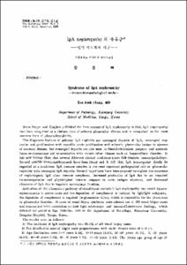
IgA Nephropathy의 증후군: 면역병리학적 연구
- Keimyung Author(s)
- Chang, Eun Sook
- Department
- Dept. of Pathology (병리학)
- Journal Title
- Keimyung Medical Journal
- Issued Date
- 1988
- Volume
- 7
- Issue
- 2
- Abstract
- Since Berger and Hinglais published the first account of IgA nephropathy in 1968, IgA nephropathy has been recognized as a distinct form of primary golmerular disease and is recognized as most common form of glomerulonephritis.
The diagnostic features of primary IgA nephritis are measngial deposits of IgA, measanial expansion and proliferation with variable acute proliferative and sclerotic glomerular lesions in absence of systemic desease, but measangial deposits are also seen in Henoch-Schonlein purpura and systemic lupus erythematosus and in association with certain other disease such as hepatobiliary disorder. It lupus erythematosus and in association with certain other disease such as hepatobiliary disorder. It has now become clear that several different clinical conditions share this common immunopathology. Several possible immnuopathogenesis have been found and it felt that IgA nephropathy should be regared as a syndrome. IgA immune complex is the most important pathogenical role in glomerulonephritis whth mesangial IgA deposits. Several hypotheses have been proposed to explain the occurence of nephritogenic IgA class immune complexes. Increased production of IgA due to and impaired immunoregulation and physiological immune response to acute antigen exposure, and decreased clearance of IgA due to impaired macrophage function.
Activation of the alternative pathway of complement cascade in IgA nephropathy can result hypocomplementemia in active cases and the deposition of complement is induced by IgG/IgM codeposits, the deposition of complement is required for glomerular injury which is responsible for the alterations in glomerular function. 41 cases of renal biopsy specimens were selected out of 133 renal biopsy cases and summerized with correlation to their light microscopic and immunofluorescence findings, which obtained the period of Sep. 1986-Oct. 1988 at the department of Pathology, Keimyung University, Dongsan Hospital, Taege, Korea.
The results were as follows:
1) The incidence of IgA nephropathy was 30.8% of all rnal biopsy cases.
2) Sex distribution showed slight male preponderance with male: female ratio of 1.05:1.
3) Age distribution were 0-9 years 2.4%, 10-19 years 19.5%, 20-29 years 46.3%, 30-39 years 14.6%, 40-49 years 7.3%, 50-59 years 7.3%, 60-69 years 2.4%. The young age group of age 10-29 years was the most prevalent.
4) The clinical symptoms were hematuria 71.8%, proteinuria(below lg/day) 4.9%, the rest above lg/day, nephrotic syndrome 24.3%, edema without nephrotic syndrome 31.7%.
5) The distributions of light microscopic findings were minimal change 19.5%, mesangial proliferation 36.6%, focal/segmental proliferation 7.3%, diffuse proliferation 17.1%, sclerosis 2.2% and crescents 2.4%.
6) Immunofluorescence study significant granular mesangial deposits of IgA and IgM 100%, IgG 75.6%, C?? 80.4%, fibrinogen 63.4%.
- Alternative Title
- Syndrome of IgA nephropathy
—Immunohistopathological study—
- Keimyung Author(s)(Kor)
- 장은숙
- Publisher
- Keimyung University School of Medicine
- Citation
- 장은숙. (1988). IgA Nephropathy의 증후군: 면역병리학적 연구. Keimyung Medical Journal, 7(2), 335–349.
- Type
- Article
- Appears in Collections:
- 2. Keimyung Medical Journal (계명의대 학술지) > 1988
1. School of Medicine (의과대학) > Dept. of Pathology (병리학)
Items in Repository are protected by copyright, with all rights reserved, unless otherwise indicated.
