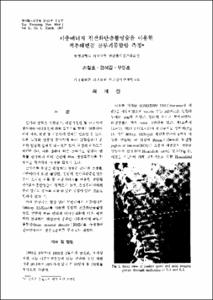
이중에너지 전산화단층촬영술을 이용한 척추해면골 골무기물함량 측정
- Keimyung Author(s)
- Sohn, Chul Ho; Woo, Young Hoon; Zeon, Seok Kil; Choi, Tae Jin
- Department
- Dept. of Medical Biophysics Engineering (의공학과)
Dept. of Radiology (영상의학)
Dept. of Nuclear Medicine (핵의학)
- Journal Title
- Keimyung Medical Journal
- Issued Date
- 1992
- Volume
- 11
- Issue
- 1
- Abstract
- Bone mineral analysis plays an important role in both detecting and managing osteoporosis and other forms of metabolic bone disease.
Dual energy quantitative computed tomography affords more accurate determintion of bone mineral density, independent of fat and water variation, and selective measurement of trabecular bone rather than integral bone provides a more sensitive means of quantifying changes in metabolic bone disease.
The bone mineral density measurements of L-1 and L-2 bodies were done in 120 Korean adults, third to eight decade, by dual energy(96,125 Kvp) quantitative CT. The elliptical region of interest was located in the anterior trabecular portion of each vertebral body.
Then the L*BMD〔(L-1+L-2 BMD)/2〕 is calculated.
The results were as follows;
The Maximum L*BMD value age group is the third decade in men, and the fourth in women.
In men, the changes of L*BMD according to age demonstrate decrease. The changes of L*BMD slowly decrease until the sixth decade(4-6%), and then rapidly decrease after the sixth decade (20%). However, in women, the changes of L*BMD accroding to age show an increase in the fourth decade, thereafter the changes of L*BMD decrease, and then rapidly decrease after the fourth decade(12-26%).
The L*BMD of healthy Korean adults is lower than of caucasian.
We propose measurement of L*BMD as a standard bone mineral density in clinical application.
- Alternative Title
- Spinal Trabecular Bone Mineral Density Measured with Dual Energy Quantitative CT
- Publisher
- Keimyung University School of Medicine
- Citation
- 손철호 et al. (1992). 이중에너지 전산화단층촬영술을 이용한 척추해면골 골무기물함량 측정. Keimyung Medical Journal, 11(1), 39–46.
- Type
- Article
Items in Repository are protected by copyright, with all rights reserved, unless otherwise indicated.
