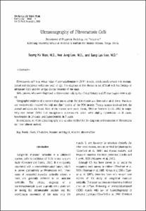
Ultrasonography of Fibromatosis Colli
- Keimyung Author(s)
- Woo, Seong Ku; Lee, Hee Jung; Lee, Sang Lak
- Department
- Dept. of Radiology (영상의학)
Dept. of Pediatrics (소아청소년학)
Institute for Medical Science (의과학 연구소)
- Journal Title
- Keimyung Medical Journal
- Issued Date
- 1996
- Volume
- 15
- Issue
- 3
- Keyword
- Neck; US studies; Neonate and Ingant; Muscles; Abnormalities
- Abstract
- Fibromatosis colli is a unique mass of sternocleidomastoid (SCM) muscle, which usually presents in neonatal period and disappears within one year of age. The diagnosis of this disease is not difficult with the finding of ultrasound (US) and the unique clinical behavior of the mass.
Sixty patients who were diagnosed as fibromatosis colli by the clinical history and US examination were studied.
Sonographic evidence of a discrete mass lesion within the SCM muscle was detected in all of them. The masses predominantly involved the mid and distal portion of the SCM muscle. Twenty masses involved both the sternal and clavicular head. Most of the masses were ovoid (n=44, 73%) or fusiform (n=12, 20%) in shape. Fifty two masses (87%) had homogeneous echo-texture, which were slightly hyperechoic in 31 cases, isoechogenic in 14 cases, and hypoechogenic in 7 cases.
In conclusion, we think ultrasonographu is a valuable method for the diagnosis and evaluation of fibromatosis colli over clinical method.
- Publisher
- Keimyung University School of Medicine
- Citation
- 우성구 et al. (1996). Ultrasonography of Fibromatosis Colli. Keimyung Medical Journal, 15(3), 219–223.
- Type
- Article
- 파일 목록
-
-
Download
 15-219.pdf
기타 데이터 / 482.47 kB / Adobe PDF
-
Items in Repository are protected by copyright, with all rights reserved, unless otherwise indicated.