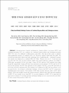
병합형 간세포암-담관암종의 임상적 및 방사선-병리학적인 양상
- Keimyung Author(s)
- Chung, Woo Jin; Jang, Byoung Kuk; Park, Kyung Sik; Cho, Kwang Bum; Hwang, Jae Seok; Ahn, Sung Hoon; Kang, Koo Jeong; Kwon, Jung Hyeok; Kang, Yu Na
- Department
- Dept. of Internal Medicine (내과학)
Dept. of Surgery (외과학)
Dept. of Radiology (영상의학)
Dept. of Pathology (병리학)
- Journal Title
- Keimyung Medical Journal
- Issued Date
- 2008
- Volume
- 27
- Issue
- 2
- Abstract
- 요 약 병합형 간세포암-담관암은 간에서 발생하는 원
발성 간암 중 드문 종양으로 임상적, 영상학적 진단만으로는 간세포암 및 담관암과 구별하기 힘든 경
우가 많으며 예후는 간세포암보다 불량한 것으로
알려져 있다. 따라서 이번 연구를 통해 병합형 간세
포암-담관암의 임상적 특징을 분석하고자 하였다.
1999년 1월부터 2007년 7월까지 계명대학교 동
산의료원에서 외과적 절제술 후 조직검사로 병합형
간세포암-담관암으로 확인된 8명의 환자들을 대상
으로 임상적, 영상학적, 병리학적 양상을 후향적으
로 분석하였다. 8명의 환자들 중 5명의 환자들에
서 혈청 αFP이 의미있게 증가하였고 5명에서
CA19-9가 증가되어 있었다. 이들 중 5명의 환자
에서 혈청 αFP및 CA19-9가 동시에 증가되어 있
어 단일 암성 병변을 예측하기는 어려웠다. 영상학
적 특징으로 5명에서 간세포암과 유사한 병변을 나
타내었고 1명은 담관암의 병변을, 2명에서는 혼재
된 양상을 보였다. 병리학적으로는 크기, 위치는 다
양하게 나타났고 간세포암 및 담관암의 특성이 8명
모두에서 동시에 관찰되었다. 변연부 및 피막, 주위
혈관 침범소견도 관찰되었다. 병합형 간세포암-담
관암의 경우 간에서 나타나는 다른 원발성 단일암
성 병변과 구별이 힘들지만 혈중의 종양 표지자가
단일암성 병변을 나타내지 못하거나 전산화 단층촬
영 소견이 종양표지자 등의 혈청학적 결과에 합당하지 않을 경우 병합형 간세포암-담관암을 염두에
두고 병기가 낮고 간기능이 양호한 상태라면 적극
적인 외과적 절제술이 필요할 것으로 생각된다.
Among primary hepatic malignancies, hepatocellular carcinoma (HCC) and
cholangiocarcinoma (CC) are common, but combined hepatocellular and cholangiocarcinoma
(HCC-CC) rare. It is difficult to differentiate each other. The aim of this study to identify
clinicopathologic feature of combined HCC-CC. Between January 1999 and July 2007, We
reviewd retrospectively clinical and radio-pathologic data of pathologically confirmed 8 patients
who initially was diagnosed as combined HCC-CC. Male : Female ratio was 6 : 2 and mean age
was 56.2 (43-74) years old. Four patients (50%) were related with liver cirrhosis. According to
TNM stage, one patient (12.5%) was stage I. Three (37.5%) and four (50%) patients were stage
II and III. respectively. Serum αFP level was elevated in seven patients (87.5%) and CA 19-9
level in five patients (62.5%) were elevated. In five patients (62.5%), serum αFP and CA19-9
level were elevated simultaneously. Five patients were diagnosed as HCC and one patients as CC
preoperativelly. According to pathologic findings, all mass had HCC and CC components
simultaneously in the masses. Four patients (50%) had vascular invasion, and 2 (25%) and 2
patients (25%) had satellite lesions and capsule invasion. Clinical course and long term prognosis
of HCC-CC are usually worse than HCC. So accurate diagnosis of HCC-CC is important for
appropriate treatment. If serologic patterns and image findings are not consistent with each other,
we must suspect the presence of HCC-CC
- Alternative Title
- Clinical and Radio-Pathologic Features of Combined Hepatocellular and Cholangiocarcinoma
- Publisher
- Keimyung University School of Medicine
- Citation
- 서혜진 et al. (2008). 병합형 간세포암-담관암종의 임상적 및 방사선-병리학적인 양상. Keimyung Medical Journal, 27(2), 125–132.
- Type
- Article
- 파일 목록
-
-
Download
 27-125.pdf
기타 데이터 / 318.02 kB / Adobe PDF
-
Items in Repository are protected by copyright, with all rights reserved, unless otherwise indicated.