KUMEL Repository
1. Journal Papers (연구논문)
1. School of Medicine (의과대학)
Dept. of Obstetrics & Gynecology (산부인과학)
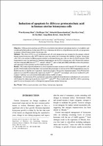
Induction of apoptosis by Hibiscus protocatechuic acid in human uterine leiomyoma cells
- Keimyung Author(s)
- Cho, Chi Heum; Shin, So Jin; Kwon, Sang Hoon; Cha, Soon Do; Song, Dae Kyu
- Journal Title
- Journal of Gynecologic Oncology
- Issued Date
- 2008
- Volume
- 19
- Issue
- 1
- Keyword
- Hibiscus protocatechuic acid; Uterus; Leiomyoma; Apoptosis
- Abstract
- Objective:Hibiscus protocatechuic acid (PCA) is a food-derived polyphenol antioxidants used as a food additive and
a traditional herbal medicine. In this study, PCA was to determine its effect on cell proliferation and cell cycle progression
in primary cultured human uterine leiomyoma cells.
Methods:The effect of PCA on cell proliferation and cell cycle progression was examined in the primary cultured
human uterine leiomyoma cells. MTT reduction assay was carried out to determine the viability of uterine leiomyoma
cells. Cell cycle analysis for Hibiscus protocatechuic acid treated leiomyoma cells was done by FACS analysis. DNA
fragmentation assay was performed to determine fragmentation rate by PCA in leiomyoma cells. Western blot analysis
was done using anti pRB, anti-p21cip1/waf1, anti-p53, anti-p27kip1, anti-cyclinE, anti CDK2 antibodies to detect the presence
and expression of these proteins in PCA treated myoma cells.
Results:PCA induced growth inhibition in a dose dependent manner, treatment with 5 mmol/L PCA blocked 80% cell
growth. FACS results showed that there was increased the percentage of cells in sub G1. DNA fragmentation assay by
ELISA was done to find the rate of apoptosis. Apoptosis took place but in a dose dependent manner. From Western blot
analysis it revealed PCA induced the expression of p21cip1/waf1 and p27kip1 increasingly and was not mediated by p53.
Caspase-7 pathway was activated and dephosphorylation of pRB took place.
Conclusion:In Conclusions, PCA, a polyphenol antioxidant, inhibited cell proliferation and induced cell cycle arrest at
sub G1 phase by enhancing the production of p21cip1/waf1 and p27kip1. These results indicate that PCA will be a promising
agent for use in chemopreventive or therapeutics against human uterine leiomyoma.
목적:본 연구는 Hibiscus protocatechuic acid (PCA)을 일차 배양된 자궁근종세포에 처리한 후 자궁근종세
포의 직접적인 증식억제 효과를 조사하고 세포주기 분석과 DNA fragmentation 분석을 통하여 세포자멸사와
의 연관성을 규명하고 그 기전을 밝히고자 세포주기관련 유전자의 발현에 대하여 조사하였다.
연구 방법:자궁근종세포를 일차배양한 후 PCA를 농도별로 처리하고 세포생존수로 증식억제효과를 관찰하고
FACS 분석 및 DNA fragmentation 분석을 통하여 세포자멸사와의 관계를 조사하였으며 Western blot
analysis 방법으로 세포주기 관련 유전자의 발현도를 측정하였다.
결과:PCA을 투여한 자궁근종세포는 농도 의존적으로 증식억제 효과가 증가하였으며 이러한 증식억제 효과는
FACS 분석 및 DNA fragmentation 분석을 통하여 괴사에 의한 것이 아니라 세포자멸사에 의한 것임을 확인하
였으며, 이러한 기전에 대하여 세포주기 관련 유전자 발현도를 측정한 결과 p21cip1/waf1과 p27kip1은 PCA의
처리 농도에 비례하여 발현도가 증가하였으며, cyclin E, A, D1 및 Bcl-2는 농도에 비례하여 발현의 감소를
확인하였다. Caspase pathway를 조사한 결과 caspase 7의 활성 증가와 pRB의 탈인산화가 관찰되었으며,
PARP 단백의 분절은 변화가 없었다.
결론:결론적으로 PCA에 의해서 자궁근종세포의 증식억제가 일어나며 이러한 현상은 세포자멸사에 의한 것이
며 그 기전은 p21cip1/waf1과 p27kip1의 발현 증가 및 pRB 인산화와 Bcl-2의 감소로 인한 세포자멸사를 확인하
였다. 이에 PCA는 세포주기와 세포자멸사에 관련된 유전자들의 발현에 영향을 미침으로써 향후 자궁근종의
치료나 예방에 있어서 효과적인 대체 치료 약물의 가능성이 있다고 사료된다.
- Alternative Title
- 자궁근종세포에 Hibiscus protocatechuic acid 투여에 의한 세포자멸사 유도
- Publisher
- School of Medicine
- Citation
- Won-Kyoung Shon et al. (2008). Induction of apoptosis by Hibiscus protocatechuic acid in human uterine leiomyoma cells. Journal of Gynecologic Oncology, 19(1), 48–56. doi: 10.3802/kjgo.2008.19.1.48
- Type
- Article
- ISSN
- 2005-0380
- 파일 목록
-
-
Download
 oak-aaa-2838.pdf
기타 데이터 / 1.1 MB / Adobe PDF
-
Items in Repository are protected by copyright, with all rights reserved, unless otherwise indicated.