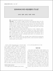
말라리아에 의한 비장침범의 CT소견
- Keimyung Author(s)
- Kwon, Jung Hyeok; Kim, Mi Jeong; Rho, Byung Hak; Lee, Mi Young
- Journal Title
- 대한영상의학회지
- Issued Date
- 2009
- Volume
- 61
- Issue
- 4
- Abstract
- Purpose: The purpose of our study was to evaluate the CT findings of malarial spleens.
Materials and Methods: We reviewed the patient records of 44 patients with malaria during a recent 3.5-year period and we selected 18 patients who underwent an abdominal CT scan. We retrospectively evaluated the CT findings of the malarial spleens and we compared then with those of a control group of 18 men. We analyzed the splenic size, whether or not there was mottled striped splenic enhancement during the arterial phase and the differences of splenic attenuation and the attenuation between the liver and spleen during the precontrast phase, the arterial phase and the portal phase between the two groups.
Results: In malarial patients, the spleen was enlarged in all cases (p < 0.001), and splenic attenuation and the degree of enhancement were significantly decreased during the precontrast phase, the arterial phase and the portal phase (p < 0.001). Loss of mottled striped enhancement during the arterial phase was seen in 11 cases (61.1%) (p < 0.001). The attenuation of the spleen was lower than that of the liver in 13 cases (72.2%) during the portal phase (p = 0.003) and in 1 case (5.6%) during the arterial phase (p = 1.000).
Conclusion: Splenomegaly, decreased splenic enhancement, the lack of mottled striped enhancement during the arterial phase and lower attenuation than that of the liver during the portal phase are helpful CT findings to diagnose the malarial spleen.
목적: 말라리아에 의한 비장침범의 CT 소견을 알아보고자 하였다.
대상과 방법: 최근 3년 6개월간 본원에서 말라리아로 진단된 44명의 환자 중에서 복부 CT를 시행한 18명의 환자를 대상으로 의무기록과 복부 CT 소견을 후향적으로 분석하였다. 비슷한 연령대의 복부 CT 소견에서 특이소견이 없는 18명을 대조군으로 하여, 말라리아 환자군과 대조군에서의 비장의 크기와 동맥기에서 비장의 불균질한 줄무늬의 조영증강 양상이 소실되는지 여부를 분석하였다. 조영증강전, 동맥기, 문맥기에서 두 군사이의 비장의 감쇠치 차이를 비교하였고, 또한 간과 비장의 감쇠치의 차이를 비교하였다.
결과: 말라리아 환자군에서 전 예에서 비장이 커져 있었으며 (p < 0.001), 조영증강전, 동맥기, 문맥기에서 통계적으로 유의하게 비장의 감쇠치가 감소하였고 (p < 0.001), 비장의 조영증강의 정도가 감소했다 (p < 0.001). 동맥기에 보이는 불균질한 줄무늬의 조영증강양상의 소실은 11예(61.1%)에서 보였으며 (p < 0.001), 문맥기에서 비장의 감쇠치가 간의 감쇠치보다 낮은 경우가 13예 (72.2%)였고 (p=0.003), 동맥기에서 비장의 감쇠치가 간의 감쇠치보다도 낮은 소견이 1예 (5.6%)였다 (p=1.000).
결론: 비장종대, 비장의 조영증강의 감소, 동맥기에서 비장의 불균질한 줄무늬의 조영증강양상의 소실, 그리고 문맥기에서 간보다 낮은 비장의 감쇠치의 소견들은 말라리아의 비장침범을 진단하는데 도움이 되는 CT 소견이다.
- Alternative Title
- CT Findings of Malarial Spleens
- Publisher
- School of Medicine
- Citation
- 장지연 et al. (2009). 말라리아에 의한 비장침범의 CT소견. 대한영상의학회지, 61(4), 241–247.
- Type
- Article
- ISSN
- 1738-2637
- Appears in Collections:
- 1. School of Medicine (의과대학) > Dept. of Preventive Medicine (예방의학)
1. School of Medicine (의과대학) > Dept. of Radiology (영상의학)
- 파일 목록
-
-
Download
 oak-bbb-02862.pdf
기타 데이터 / 517.69 kB / Adobe PDF
-
Items in Repository are protected by copyright, with all rights reserved, unless otherwise indicated.