KUMEL Repository
1. Journal Papers (연구논문)
1. School of Medicine (의과대학)
Dept. of Obstetrics & Gynecology (산부인과학)
임신 주수와 조기양막파수 유무에 따른 양막 두께 변화 및 병리조직 소견의 비교연구
- Keimyung Author(s)
- Park, Joon Cheol; Yoon, Sung Do
- Department
- Dept. of Obstetrics & Gynecology (산부인과학)
- Journal Title
- 대한산부인과학회잡지
- Issued Date
- 2003
- Volume
- 46
- Issue
- 7
- Abstract
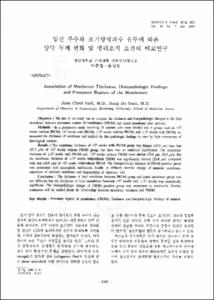
- Objective : The aim of our study was to compare the thickness and histopathologic changes in the fetal membrane between premature rupture of membranes (PROM) and intact membrane after delivery. Methods : In a prospective study involving 31 patients who were divided into 4 groups such as <37 weeks without PROM, <37 weeks with PROM, ≥37 weeks without PROM, and ≥37 weeks with PROM, we measured the thickness of membrane and studied the histopathologic findings in vitro by light microscopy of histological sections. Results : The membrane thickness of <37 weeks with PROM group was thinner (35.9 ㎛) than that (42.3 ㎛) of <37 weeks without PROM group, but there was no statistical significance. The membrane thickness of ≥37 weeks with PROM and ≥37 weeks without PROM were similar (25.6 ㎛, 26.0 ㎛). But the membrane thickness of ≥37 weeks with/without PROM was significantly thinner (25.8 ㎛) compared with that (38.9 ㎛) of <37 weeks with/without PROM. The histopathologic features of PROM positive group was amnionitis with neutrophilic infiltration, focally or diffusely necrotic change of amniotic membrane, separation of amniotic membrane and degeneration of chorionic villi. Conclusion : The thickness of fetal membrane between PROM group and intact membrane group was not different but the thickness of fetal membrane between <37 weeks and ≥37 weeks was statistically significant. The histopathologic change of PROM positive group was prominent as amnionitis. Further evaluation will be needed about the relationship between membrane thickness and PROM.
Key word : Premature rupture of membranes (PROM), Thickness and histopathologic findings of amnion
목적 : 조기 파수된 산모의 분만 후 얻은 태아막을 이용하여 양막의 두께를 측정하여 같은 임신 주수의 대조군과 상호 비교하여 양막의 두께와 조기 양막 파수와의 연관관계를 보고자 하였고 융모양막의 조직학적 병리소견을 관찰하였다. 연구 방법 : 31명의 산모에서 분만 후 얻은 융모양막의 두께 및 병리학적 검사를 시행하였다. 이중 18명은 조기 양막 파수로 진단되어 입원한 산모였다. 이들을 다시 주수에 따라 구분하여 4개의 군으로 세분하여 37주 미만의 양수 파수가 없는 군, 37주 미만의 조기 양막 파수된 군, 37주 이상의 양막 파수가 없는 군, 37주 이상의 조기 양막 파수된 군으로 구분하였다. 결과 : 37주 미만의 조기 양막 파수된 군에서 양막 두께는 35.9 ㎛로 37주 미만의 양수 파수가 없는 군의 42.3㎛보다 얇았으나 Mann-Whitney test상 P-value 0.116로 통계학적 유의성은 없었다. 그리고 37주 이상의 조기양막 파수된 군은 25.6㎛으로 같은 주수의 양막 파수가 없는 군의 26.0㎛로 별다른 차이가 없었다. 환자군을 조기 양막 파수 여부에 따라 두 군으로 구분시에도 31.1㎛와 33.1㎛으로 조기 양막 파수 유무에 따른 차이 25.8㎛로 37주 미만의 38.9㎛에 비하여 유의하게 얇게 측정되었다. 결론 : 임신 만기에 가까울수록 양막의 두께가 유의하게 감소하였으나, 조기 양막 파수된 환자에서 태아막의 두께 차이는 통계학적 유의성이 없었다. 그러나 조기 양막 파수된 경우 백혈구 침윤이나 양막의 분리, 융모의 변성이나 괴사, 혹은 기저막의 초자양 변성 등이 현저하였다.
중심단어 : 조기 양막 파수, 양막의 두께, 조직학적 소견
- Alternative Title
- Association of Membrane Thickness, Histopathologic Findings and Premature Rupture of the Membranes
- Publisher
- School of Medicine
- Citation
- 박준철 et al. (2003). 임신 주수와 조기양막파수 유무에 따른 양막 두께 변화 및 병리조직 소견의 비교연구. 대한산부인과학회잡지, 46(7), 1385–1390.
- Type
- Article
- ISSN
- 0494-4755
- Appears in Collections:
- 1. School of Medicine (의과대학) > Dept. of Obstetrics & Gynecology (산부인과학)
- 파일 목록
-
-
Download
 oak-bbb-1527.pdf
기타 데이터 / 8.39 MB / Adobe PDF
-
Items in Repository are protected by copyright, with all rights reserved, unless otherwise indicated.