KUMEL Repository
1. Journal Papers (연구논문)
1. School of Medicine (의과대학)
Dept. of Obstetrics & Gynecology (산부인과학)
자궁 근종 세포에서 ATP-민감성 칼륨 통로의 동정
- Keimyung Author(s)
- Cha, Soon Do; Cho, Chi Heum; Song, Dae Kyu
- Journal Title
- 대한산부인과학회잡지
- Issued Date
- 2003
- Volume
- 46
- Issue
- 12
- Abstract
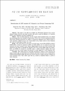
- Objective : The purpose of this study was to identify the ATP-sensitive potassium (KATP) channel subtypes in uterine leiomyoma and normal myometrial cells, and to compare the difference in its expression between uterine leiomyoma and normal myometrial cells.
Methods : Fresh ten uterine leiomyomas and their adjacent normal myometrial tissues were obtained from hysterectomies that were conducted on benign diseases. With the use of reverse transcription-polymerase chain reaction (RT-PCR) and Western blot analysis, we analysed the expression of KATP channel subtypes in tissues and primary cultured leiomyoma cells. To demonstrate the KATP channel activity in uterine leiomyoma cells, whole cell patch-clamp recordings were made.
Results : The uterine leiomyoma and normal myometrial tissues contained Kir6.1/SUR2B and Kir6.2/ SUR2B mRNAs, although the expression of SUR1 and SUR2A mRNAs were not expressed in the uterine leiomyoma and normal myometrial tissues. Primary cultured uterine leiomyoma and normal myometrial cells demonstrated same patterns. To determine the expression levels of KATP channel subunits, high levels of Kir6.1, Kir6.2, and SUR2B were detected by western-blot analysis in uterine leiomyoma compared with normal myometrial tissues. KATP channel currents were increased by estrogen application in uterine leiomyoma cells.
Conclusion : These studies provide new knowledge concerning KATP channels in uterine leiomyoma and normal myomtrial cells. we demonstrated uterine leiomyoma and normal myometrial tissues contained Kir6.1/ SUR2B and Kir6.2/SUR2B subunits and showed an increased expression in uterine leiomyoma compared with normal myometrial tissues. These results suggest that KATP channels are important elements in growth of uterine leiomyoma.
목적 : 본 연구는 지금까지 자궁 근종의 KATP channel 아형에 관한 연구가 없어, 자궁 근종이나 정상 자궁근에서의 KATP channel의 존재와 아형의 종류를 확인하고, 또한 발현의 차이를 확인하고자 하였다.
연구 방법 : 자궁근종으로 전자궁 적출술을 시행하는 10예의 환자를 대상으로 자궁 근종과 인접한 정상 자궁 조직을 얻어 실험하였고, 또한 일차 세포배양을 하였다. 자궁내막의 주기는 증식기 5예와 분비기 5예로 하였다. 조직과 일차배양세포에서 KATP channel 아형의 RT-PCR, western-blot 분석을 하였으며, KATP 통로의 전기적인 활성도를 측정하기 위하여 patch clamp 기법의 whole-cell mode를 사용하였다.
결과 : RT-PCR을 통한 KATP channel의 아형의 검사에서 Kir6.1, Kir6.2, SUR 2B는 자궁 근종과 정상 자궁근에서 모두 발현이 있었으나, SUR1과 SUR2A의 발현은 보이지 않았고, 세포를 일차 배양한 세포에서도 동일한 결과를 보였다. Western Blot 분석에서는 Kir6.1, Kir6.2, SUR 2는 자궁 근종과 정상 자궁 근에서 모두 발현이 있었고, SUR1의 발현은 보이지 않았다. KATP channel의 아형의 발현 양상에서는 SUR2, Kir6.1과 Kir6.2는 자궁 근종에서 자궁 근조직보다 높은 발현을 보였다. whole-cell mode에서 K+ 이온이 야기하는 내향성 전류가 관찰되었으며, 에스트로겐에 노출시 초기 신호전달에 세포막의 KATP channel을 통한 전류의 증가를 관찰하였다.
결론 : 본 연구에서는 KATP channel 아형을 동정하여 자궁 근종과 정상 자궁 근에서 Kir6.1/SUR2B와 Kir6.2/SUR2B가 존재하는 것을 밝혔으며, 또한 자궁 근종 세포에서 KATP channel의 발현이 증가함을 확인하였다. 이 결과를 이용하여 자궁 근종 세포 증식의 조절 기전을 밝히는데 도움이 되리라 생각되며, 향후 KATP channel 이온 통로 조절 인자를 이용한 자궁 근종의 치료에 이용할 수 있으리라 사료된다.
중심단어 : 자궁근종, KATP channel
- Alternative Title
- Identification of ATP-sensitive K+ Channel in an Uterine Leiomyoma Cell
- Publisher
- School of Medicine
- Citation
- 차순도 et al. (2003). 자궁 근종 세포에서 ATP-민감성 칼륨 통로의 동정. 대한산부인과학회잡지, 46(12), 2380–2385.
- Type
- Article
- ISSN
- 0494-4755
- 파일 목록
-
-
Download
 oak-bbb-1533.pdf
기타 데이터 / 371.87 kB / Adobe PDF
-
Items in Repository are protected by copyright, with all rights reserved, unless otherwise indicated.