KUMEL Repository
1. Journal Papers (연구논문)
1. School of Medicine (의과대학)
Dept. of Obstetrics & Gynecology (산부인과학)
Histone Deacetylase 억제제로 유도된 자궁내막암 세포의 세포증식 억제와 세포자멸사 기전연구
- Keimyung Author(s)
- Shin, So Jin; Kwon, Sang Hoon; Cho, Chi Heum; Cha, Soon Do
- Department
- Dept. of Obstetrics & Gynecology (산부인과학)
- Journal Title
- 대한산부인과학회지
- Issued Date
- 2009
- Volume
- 52
- Issue
- 9
- Abstract
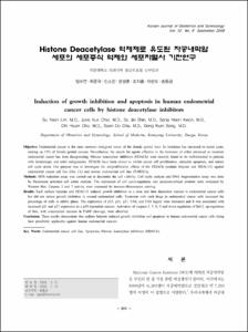
- Objective: Endometrial cancer is the most common malignant tumor of the female genital tract. Its incidence has increased in recent years, making up 13% of female genital cancers. Nevertheless, the search for agents effective in the treatment of either advanced or recurrent endometrial cancer has been disappointing. Histone deacetylase inhibitors (HDACIs) were recently found to be well-tolerated in patients with hematologic and solid malignancies. HDACIs have been shown to inhibit cancer cell proliferation, stimulate apoptosis, and induce cell cycle arrest. Our purpose was to investigate the antiproliferative effects of the HDACIs (sodium butyrate and HDAC-I1) against endometrial cancer cell line (Hec 1A) and normal endometrial cell line (T-HESCs).
Methods: MTS reduction assay was carried out to determine the cell viability. Cell cycle analysis and DNA fragmentation assay was done by fluorescent activated cell sorter analysis. The expression of cell cycle-regulatory and apoptosis-related proteins were evaluated by Western blot. Caspase 3 and 7 activity were measured by immuno-flouorescent staining.
Results: Each sodium butyrate and HDAC-I1 induced growth inhibition in a dose and time dependent manner in endometrial cancer cells but did not induce growth inhibition in normal endometrial cells. Treatment with each drugs in endometrial cancer cells increased the percentage of cells in subG1 phase. The expression of p53, p21, p27, FAS, and FAS legand were increased and it was associated with increased p21 and p27 expression in a p53-dependent manner. Activation of caspase-3, 7, 8, 9 and down-regulation of Bcl-2, up-regulation of Bax, with concomitant increase in PARP cleavage, were observed.
Conclusion: These results demonstrate that sodium butyrate induced growth inhibition and apoptosis in human endometrial cancer cells rising their possibility applicable against human endometrial cancers.
목적: 자궁내막암은 최근 급속한 경제 성장에 따른 생활수준의 향상으로 말미암아 평균 수명이 연장되고 점차 발생 빈도가 증가하고 있는 중요한 부인암의 하나이다. 자궁내막암 환자의 70~80%는 암병기 제 1기에 속하며 생존율이 비교적 양호한
경과를 보이지만 진행암 및 재발암의 경우에는 다른 악성 부인과 종양과 마찬가지로 불량한 예후를 보이며, 진행된 암에서의 치료는 지금까지 적절한 약물치료가 없다. Histone Deacetylase (HDAC) 억제제는 악성화되는 과정에 있는 세포를 복구하는 능력을 가진 새로운 항암치료제이다. 그렇지만 HDACIs의 구체적인 작용기전에 대해서는 아직 명확히 밝혀지고 있지 않다. 그러므로 본 연구자는 천연 물질인 NaB와 합성된 HDAC-I1을 이용하여 자궁내막암 세포주인 Hec 1A 세포주와 정상자궁 내막 세포주인 THESCs 세포에 어떤 영향을 미치는지 알아보고 세포에 미치는 영향을 분석하여 치료에 유용한지를 알아
보고자 하였다.
연구 방법: NaB와 HDAC-I1 각각 투여 후 자궁내막암 세포와 정상 자궁 내막세포에서의 세포생존율을 알아보기 위해 MTS분석을 이용하였고, 세포 주기 회로 및 세포자멸사에 관계하는 유전자의 발현을 보기 위해 Western blot analysis를 시행하였다. 세포주기분석은 유세포분석기를 이용하였으며 세포자멸사를 확인하기 위해 DNA fragmentation 방법을 이용하였다. Caspase의 활성도를 확인하기 위해 면역형광 염색을 시행하였다.
결과: 자궁내막암 세포에 NaB와 HDAC-I1을 농도별로 처리하여 24시간 후의 결과에서 농도 및 시간 의존적으로 증식억제의 증가를 보였으나 정상 세포에서는 성장 억제 효과는 보이지 않았다. 세포 주기분석에서 자궁내막암 세포에 투여 시 subG1주기의 지연이 농도가 증가할수록 증가하였으나, 정상 자궁내막 세포에서는 세포주기의 변화가 없었다. 세포자멸사의 증거인 DNA 분절 검사를 시행하여본 결과 자궁내막암 세포가 NaB에 의해 농도가 증가할수록 세포분절현상이 증가되는 것을 확인하였다. p53, p21, p27, FAS와 FAS ligand 단백은 농도가 증가할수록 발현이 증가하였고, CDK2와 cyclin A의 발현 감소를
확인하였으며, 미토콘드리아에서 세포자멸사에 관계하는 Bcl2는 감소하였으나 Bax 단백은 반대로 발현 증가를 보였다. 비활성 형태의 pro-caspase 3과 8 단백질의 양적 감소, caspase 9의 활성과 PARP cleavage의 증가되어 세포자멸사로 유도되는 것을 알았다. caspase 7과 3의 활성도 검사에서도 면역형광으로 활성이 증가하는 것을 관찰하였다.
결론: 이상의 결과를 통해 NaB와 HDAC-I1이 자궁내막암 세포에 내인성과 외인성의 세포자멸사의 경로를 거쳐 세포주기와 세포자멸사에 관련된 유전자들의 발현에 영향을 미침으로써 자궁내막암 세포의 증식을 억제하여 결과적으로 세포자멸사에 이르게 함을 알았으며, 향후 자궁내막암의 치료 약물로서의 가능성을 보인다고 생각된다.
- Alternative Title
- Induction of growth inhibition and apoptosis in human endometrial cancer cells by histone deacetylase inhibitors
- Publisher
- School of Medicine
- Citation
- 임수연 et al. (2009). Histone Deacetylase 억제제로 유도된 자궁내막암 세포의 세포증식 억제와 세포자멸사 기전연구. 대한산부인과학회지, 52(9), 911–919.
- Type
- Article
- ISSN
- 1738-5628
- Appears in Collections:
- 1. School of Medicine (의과대학) > Dept. of Obstetrics & Gynecology (산부인과학)
- 파일 목록
-
-
Download
 oak-bbb-1560.pdf
기타 데이터 / 1.44 MB / Adobe PDF
-
Items in Repository are protected by copyright, with all rights reserved, unless otherwise indicated.