KUMEL Repository
1. Journal Papers (연구논문)
1. School of Medicine (의과대학)
Dept. of Neurosurgery (신경외과학)
백서의 실험적 뇌지주막하 출혈에서 Nimodipine이 대뇌조직의 Leukotriene C₄ 함량에 미치는 영향
- Keimyung Author(s)
- Yim, Man Bin; Lee, Jang Chull; Son, Eun Ik; Kim, Dong Won; Kim, In Hong; Lee, In Kyu
- Journal Title
- 대한신경외과학회지
- Issued Date
- 1992
- Volume
- 21
- Issue
- 9
- Abstract
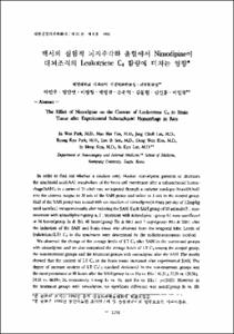
- In order to find out whether a calcium entry blocker, nimodipine, prevents or decreases the arachnoid acid(AA) metabolism of the brain cell membrane after a subarachnoid hemorrhage(SAH), in a series of 35 adult rats, we injected through a catheter autologus blood(0.3㎖) into the cisterna magna in 30 cats of the SAH group and saline in 5 rats in the control group. Half of the SAH group was treated with an injection of nimodipine(4 times per day of 1.2㎎/㎏ until sacrifice) intraperitoneally after inducing the SAH. Each SAH group of 10 animals(5 : non-treatment with nimodipine=group a. 5 : treatment with nimodipine=group a. 5 : treatment with nimodipine=group b) were sacrifieced at 24 hours(group Ia & Ib), 48 hours(group Ⅱa & Ⅱb) and 7 days(group Ⅲa & Ⅲb) after the induction of the SAH and brain tissue was obtained from the temporal lobe. Levels of leukotriene(LT) C₄ in the specimens were determined by the radioimmunoassay method.
We observed the change of the average levels of LT C₄ after SAH in the nontreated groups with nimodipine, and we also compaired the average levels of LT C₄ among the control group, the non-treatment groups and the treatment groups with nimodipine after the SAH. The results showed that the content of LT C₄ in the brain tissue increased after experimental SAH. The degree of increase content of LT C₄ (±standard deviation) in the non-treatment groups was the most prominent at 48 hours after the SAH(group Ia vs. Ⅱa vs. Ⅲ a : 61.31±22.28 vs. 120.38±24.18 vs. 66.84±28, respectively. Group Ia vs. Ⅱa, and Ⅱa vs. Ⅲ a ; p<0.05). However, in the treatment groups with nimodipine, no significant difference was noted(group Ib vs. Ⅱb vs. Ⅲ b : 49.19±8.19 vs. 42.04±14.66 vs. 47.19±17.84, respectively). Levels of LT C₄ in the treatment groups were lower than those of the non-treatment groups, especially at 48 hours after the SAH(group Ⅱa vs. Ⅱb : 120.38±24.18 vs. 42.04±14.66, respectively, p<0.05).
This study showed that nimodipine suppressed the release of LT C₄ in brain tissue after the SAH and its protective effect was the most prominent at 48 hours after the SAH. We conclude that nimodipine, aside from its vascular effect, may expert a protective role against the damage of neurons after the SAH with a decrease in the release of the lipoxygenase pathway metabolites of AA.
본 교실에서는 뇌동맥류 파열에 기인한 뇌지주막하 출혈 환자에서 nimodipine이 뇌혈관 연축에 기인한 뇌허혈 상태를 호전시키는 기전의 하나로서, 뇌세포외 칼슘이온의 뇌세포내 이동을 차단시켜 뇌세포막의 AA 대사를 저하시키므로써 뇌세포 손상에 관여하는 AA 대사산물들의 생성을 억제하여 뇌허혈 상태를 호전시키는지 여부를 알아보고자 실험을 시행하였다. 실험 동물은 백서를 사용하였고 뇌지주막하 출혈은 대조에 도관을 삽입하여 자가 혈로 유도하였다. AA 대사산물은 측두엽에서 채취한 대뇌조직에서 방사선면역측정법으로 LT C₄를 측정하였고, nimodipine은 복강내로 주입하여 치료하였다. 결과는 뇌지주막하 출혈후 대뇌조직에서도 AA의 lipoxygenase 대사산물, 즉 LT C₄는 증가되었으며, 특히 조기인 48시간에 가장 현저하였다. Nimodipine치료는 전 실험기간을 통하여 AA의 lipoxygenase 대사산물인 LT C₄의 생성을 억제하였고 특히 48시간군에서 현저하였다. 따라서 nimodipine은 뇌지주막하 출혈에서 뇌혈관 연축에 기인한 뇌허혈 증상을 호전시키는 기전으로 미세뇌혈관을 확장시켜 뇌혈류를 개선시키는 것이외에 뇌세포막의 AA 대사를 억제하여 뇌세포의 손상을 방지하는 것도 하나의 기전이 될 것으로 추정하며, 아울러 nimodipine의 LT C₄생성에 대한 억제효과는 48시간군에서 가장 현저하였으므로 뇌지주막하 출혈환자에서 nimodipine을 사용시 출혈후 조기에 사용하는 것이 좋다는 사실도 뒷받침한다 하겠다.
- Alternative Title
- The Effect of Nimodipine on the Content of Leukotriene C₄ in Brain Tissue after Experimental Subarachnoid Hemorrhage in Rats
- Publisher
- School of Medicine
- Citation
- 박인우 et al. (1992). 백서의 실험적 뇌지주막하 출혈에서 Nimodipine이 대뇌조직의 Leukotriene C₄ 함량에 미치는 영향. 대한신경외과학회지, 21(9), 1138–1146.
- Type
- Article
- ISSN
- 1225-8245
- Appears in Collections:
- 1. School of Medicine (의과대학) > Dept. of Internal Medicine (내과학)
1. School of Medicine (의과대학) > Dept. of Neurosurgery (신경외과학)
- 파일 목록
-
-
Download
 oak-bbb-1626.pdf
기타 데이터 / 567.43 kB / Adobe PDF
-
Items in Repository are protected by copyright, with all rights reserved, unless otherwise indicated.