KUMEL Repository
1. Journal Papers (연구논문)
1. School of Medicine (의과대학)
Dept. of Neurosurgery (신경외과학)
백서의 중대뇌동맥 폐쇄로 유발시킨 뇌경색의 범위 측정과 경시적 변동
- Keimyung Author(s)
- Kim, In Soo; Yim, Man Bin; Lee, Jang Chull; Son, Eun Ik; Kim, Dong Won; Kim, In Hong; Kwon, Kun Young
- Journal Title
- 대한신경외과학회지
- Issued Date
- 1992
- Volume
- 21
- Issue
- 1
- Abstract
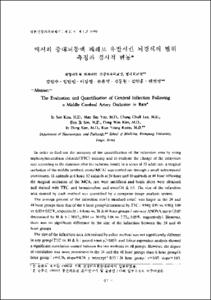
- In order to find out the accuracy of the quantification of the infarction area by using triphenyltetrazolium chloride(TTC) staining and to evaluate the change of the infarction size according to the duration after the ischemic insult, in a series of 33 adult rats, a surgical occlusion of the middle cerebral artery(MCA) was carried out through a small subtemporal craniotomy. 11 animals at 6 hour, 12 animals at 24 hour and 10 animals at 48 hour following the surgical occlusion of the MCA, rats were sacrificed and brain slices were obtained and stained with TTC, and hematoxyline and eosin(H & E). The size of the infarction area stained by each method was quantified by a computer image analysis system. The average percent of the infarction size(± standard error) was larger in the 24 and 48 hour groups than that of the 6 hour group(determined by TTC : 9.94±0.97 vs. 9.98±1.08 vs. 6.83±0.82%, respectively : 6 hour vs. 24 & 48 hour groups; one-way ANOVA test p<0.05 determined by H & E : 10.02±0.94 vs. 10.02±1.06 vs. 7.73±0.85%, respectively). However, there was no significant difference in the size of the infarction between the 24 and 48 hour groups. The size of the infarction area determined by either method was not significantly different in any group(TTC vs. H & E : paired t-test p>0.05), and linear regression analysis showed a significant correlation existed between the two methods in all groups. However, the degree of correlation was more prominent in the 24 and the 48 hour groups than 6 hour group(6 hour group : r=0.76, slope=0.78, y intercept=0.55;24 hour group : r=0.97, slope=1.03, y intercept=-0.78;48 hour group : r=0.98, slope=0.94, y intercept=0.42). From this study it is concluded that : 1) the evolution of the infarction size continues up to 24 hours following the arterial occlusion, and thereafter, the change of the infarction size is minimal in the rat. This data suggests that it is sufficient to evaluate the change of the infarction size up to 24 hours following the ischemic insult in the experimental study of ischemia in the rat. 2) the detection and the quantification of the cerebral infarction by using TTC staining is a reliable method after 24 hours following the ischemic insult. However, in the earlier period than 24 hours following the ischemic insult, staining with TTC coupled with histopathological H & E staining will add to the accuracy in the obtaining the quantity of the cerebral infarction in the rat.
본교실에서는 33마리의 백서를 실험동물로 하여 측두하 접근으로 중대뇌동맥을 폐쇄하여 뇌경색을 유발하고, 뇌경색의 범위측정을 뇌허혈 유발후 6시간, 24시간 및 48시간에 TTC 염색과 병리조직학적인 H & E 염색으로 하였다. 뇌허혈 유발후 뇌경색 범위는 6시간군에 비하여 24시간 및 48시간군에서 보다 현저히 증가하였으나 24시간 군 및 48시간 군간에는 별 차이가 없었다. 따라서 본 실험을 통해서 백서를 실험동물로 하여 뇌경색에 대한 실험을 시행시 뇌경색의 크기를 기준하여 실험 결과를 판정할 경우 뇌허혈 유발후 24시간까지만 실험 기간으로 하여도 무방할 것이라는 점을 알았다. TTC 염색과 H & E 염색의 상관 관계에서는 모든 실험 기간에서 높은 상관 관계를 보이나 6시간군에서는 상대적으로 낮으므로, 뇌허혈 유발후 조기, 즉 24시간 이내에는 병리조직학적 방법을 병행하여 시행하는 것이 더욱 정확성을 기할 것으로 생각되었다
- Alternative Title
- The Evaluation and Quantification of Cerebral Infarction Following a Middle Cerebral Artery Occlusion in Rats
- Publisher
- School of Medicine
- Citation
- 김인수 et al. (1992). 백서의 중대뇌동맥 폐쇄로 유발시킨 뇌경색의 범위 측정과 경시적 변동. 대한신경외과학회지, 21(1), 97–108.
- Type
- Article
- ISSN
- 1225-8245
- Appears in Collections:
- 1. School of Medicine (의과대학) > Dept. of Neurosurgery (신경외과학)
1. School of Medicine (의과대학) > Dept. of Pathology (병리학)
- 파일 목록
-
-
Download
 oak-bbb-1628.pdf
기타 데이터 / 562.5 kB / Adobe PDF
-
Items in Repository are protected by copyright, with all rights reserved, unless otherwise indicated.