KUMEL Repository
1. Journal Papers (연구논문)
1. School of Medicine (의과대학)
Dept. of Plastic Surgery (성형외과학)
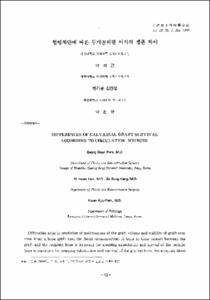
혈행차단에 따른 두개골외판 이식의 생존차이
- Keimyung Author(s)
- Han, Ki Hwan; Kang, Jin Sung; Park, Kwan Kyu
- Journal Title
- 대한성형외과학회지
- Issued Date
- 1993
- Volume
- 20
- Issue
- 1
- Abstract
- Difficulties arise in prediction of maintenance of the graft volume and viability of graft over time when a bone graft used for facial reconstruction. A bone-to-bone contact between the graft and the recipient bone is imporant for creeping substitution and survial of the recipient bone is important for creeping substitution and of the grafted bone. An adequate blood supply is also essential to ensure the survival of any live cells of the surface of the graft.
Our study was designed to determine which one is an important factor viability of the grafted bone in Korean adult dogs : a bone-to-bone contact or overlying soft tissue. Blocks of outer table of the parietal bone were placed at the maxillae subperiostially in 4 different ways : bone-to-bone contact groups(groups Ⅰand Ⅱ)with placing a silicone membrane over the grafted bone and soft tissue contact groups(groups Ⅲand Ⅳ)with placing a silicone sheet between the grafted bone and the recipient. In groups Ⅰand Ⅲ, the cancellous surfaces of the parietal bone was placed on the recipient and the cortical surfaces were placed on the recipient in groups Ⅱ and Ⅳ. Caliper techniques were used to study the rates of volume maintenance of the grafts at 6, 12, and 20 weeks after bone grafting. The volumes of the living bone were quantified microscopically using a modified point-countiong technique.
The volume is reduced in a similar rate with time in all groups. At 6 week, living bone cells increased in soft tissue contact groups Ⅲ and Ⅳ however, and increased in bone to bone contact groups Ⅰand Ⅱ at 12 and 20 weeks. there were osteoblastic proliferation and laminated mature bones in group Ⅰand Ⅱ. But osteoclasts and their associated osteolytic changes were still seen near the silicone membrane in group Ⅲ and Ⅳ, which may imply a continuing resorptive process with time.
In summary, revascularization from the overlying soft tissue is important for the graft survival in early stage of the bone grafting while bone-to-bone contact may be essential in a later stage.
저자들은 골이식의 생존에 수혜골과의 적당한 접합이 중요한 지 아니면 이식골을 더는 연조직이 중요한 지를 연구하고자 동물실험을 실시하였다. 성숙견 12마리를 두개골 외판의 해면질골면과 피질골면 중 어느 한 면은 실리콘판으로 덮어 싸서 혈관화를 차단한 뒤 다른 한 면이 각각 수혜골 및 연조직과 접합하도록 4군으로 나누어 이식한 다음 6주, 12주 및 20주에 육안 및 광학현미경으로 관찰하였을 때 다음과 같은 지견을 얻을 수 있었다.
1. 연조직으로부터 들어오는 혈행을 차단한 군이나 수혜골과의 골합착을 차단한 군이나 시간경과에 따라 별 차이 없이 비슷하게 골용적이 감소하였다. 그러나 조직학적으로는 유의한 차이를 나타낸 것을 볼 때 이식골이 잘 생존하려면 수혜골에 잘 접합시키는 것이 중요할 것으로 생각된다.
2. 이식 초기에는 수혜골에의 합착보다는 연조직으로부터의 혈행이 골생존에 더 중요하지만 시간이 흐름에 따라 수혜골과의 합착이 잘 이루어져야 골흡수가 적고 골부가가 많아져서 골생존이 좋을 것으로 생각된다.
3. 해면질골면 또는 피질골면 중 어느 쪽을 수혜골에 접하거나 연조직에 접하도록 이식하더라도 즉 동일한 수혜부에서는 골흡수와 골생존에 유의한 차이는 없을 것으로 생각된다.
4. 수혜골로부터 골합착 및 혈행을 차단한 경우 골부가 과정 때인 술후 20주에도 골파괴소견 및 파골세포가 관찰되는 것으로 보아 장시간 뒤에는 골흡수가 좀 더 일어날 것으로 예상된다.
이상을 종합해 보면 골생존에는 수혜부 연조직으로부터의 혈행이나 수혜골과의 골합착이 모두 비슷하게 중요하지만 장기적으로 볼 때 수혜골과 골합착이 잘 되도록 이식하는 것이 골흡수가 적을 것으로 판단된다.
- Alternative Title
- DIFFERENCES OF CALVARIAL GRAFT SURVIVAL ACCORDING TO CIRCULATION SOURCES
- Publisher
- School of Medicine
- Citation
- 박성근 et al. (1993). 혈행차단에 따른 두개골외판 이식의 생존차이. 대한성형외과학회지, 20(1), 61–72.
- Type
- Article
- ISSN
- 1015-6402
- Appears in Collections:
- 1. School of Medicine (의과대학) > Dept. of Pathology (병리학)
1. School of Medicine (의과대학) > Dept. of Plastic Surgery (성형외과학)
- 파일 목록
-
-
Download
 oak-bbb-1631.pdf
기타 데이터 / 1.64 MB / Adobe PDF
-
Items in Repository are protected by copyright, with all rights reserved, unless otherwise indicated.