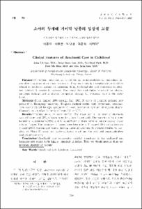
소아의 두개내 거미막 낭종의 임상적 고찰
- Keimyung Author(s)
- Kim, Joon Sik; Lee, Hee Jung
- Journal Title
- 대한소아신경학회지
- Issued Date
- 2002
- Volume
- 10
- Issue
- 2
- Abstract
- Purpose: Intracranial arachnoid cysts are benign neurodevelopmental anomalies that are often diagnosed in childhood incidently. They are clinically asymptomatic or could be related to headache, seizure, devlopmental delay, hydrocephalus and sometimes to attention deficient hyperactivity disorder. This study was undertaken to review the clinical, radiologic findings and to discuss therapeutic strategy for arachnoid cyst in the childhood.
Methods: From August 1996 through July 2002, 26 cases of pediatric patients hospitalized in Keimyung university, Dongsan medical center with intracranial arachnoid cyst were analyzed for age, symptoms of onset, location of cyst and therapeutic detalis. Diagnosis was ratified by using of brain CT or MRI.
Results: Twenty-six cases were studied. The mean age at the time of diagnosis was 5.2 years and 31% of them were less than 2 years old. The majority of cyst were located in supratentorial(88%) and 16 cases(61%) of them were on middle cranial fossa/ sylvian fissure. The symptoms of onset were headache in 11 cases(42%), convulsions on 6 cases(23%), trauma and others. Among these 26 children, 18 children treated by surgery, in which 10 cases had cysto-peritoneal shunt and the rest had marsupialization and excision of cyst.
Conclusion: Arachnoid cyst represented variable symptoms in the childhood and incidence rate seems to be higher, especially in infant. Thus we should provide them appropriate strategy of therapy.
목 적:거미막 낭종은 구조적인 중복 또는 분열로 인하여 거미막내에 뇌척수액과 유사한 체액이 고이는 질환으로 소아기에 우연히 발견되는 발육이상으로 알려져 있으나 발생부위에 따라 유발되는 증상이 다를 뿐 아니라 간질, 경막하 혈종, 뇌수종, 주의력 결핍 과잉 행동장애 및 정신 운동 발달지연의 원인이 된다는 보고가 증가하는 추세이다. 대부분의 경우 보존적 요법으로 치료가 충분하다는 주장과는 달리 적극적인 수술적 치료가 도움이 된다는 주장도 있다. 이러한 거미막 낭종의 임상적 특성을 알아보고 치료의 결과에 도움을 주고자 본 연구를 시행하였다.
방 법:1996년 8월부터 2002년 7월까지 6년간 계명대학교 의과대학 동산의료원에서 두부전산화찰영 또는 자기공명영상을 시행하여 거미막 낭종이 발견되었던 26명의 환아를 대상으로 연령, 호발 증상, 거미막 낭종의 위치, 수술 여부에 대하여 후향적으로 분석하였다.
결 과:전체 26명의 환아중 남아가 14명(54%), 여아는 12명(46%)으로 남녀비는 1.2:1로 남아에서 조금 많았다. 진단 당시의 나이는 평균적으로 5.2세였다. 2세 이전에서의 발생빈도는 8례로서 전체의 31%에 해당하였다. 거미막 낭종의 발생 위치는 전체 26례 중 14례(54%)에서 좌측에 발생하였고 7례(27%)에서 우측, 그리고 5례(19%)는 양측성으로 발생하였다. 낭종의 위치는 23례(88%)가 천막상부(supratentorial)에 위치하고 나머지 3례(12%)많이 천막하부에 위치하였다. 천막상부에 위치한 낭종의 16례(61%)가 중두와에 또는 실비안열(sylvian fissure)에 위치하여 가장 많았으며 이외에도 안장상부(suprasellar) 4례(15%)가 있었고 기타 6례로 전두부에 2례, 두정부에 1례, 그리고 천막하부에는 후두와에 3례가 발견되었다. 자기공명영상을 시행하게 된 초발증상으로는 두통 11례(42%)로 가장 많았으며 경련 6례(23%), 외상 4례(15%), 대두증 등이었으며 그외에도 신생아가사, 무월경증이 동반 된 내분비 장애 등이 있었다. 전단된 26례의 환아를 대상으로 치료는 18례(69%)에서 수술을 시행되었으며, 수술이 시행된 경우의 10례(56%)에서는 낭종 복강간 단락술을 시행하였고 수술을 시행하지 않은 경우는 질병의 경과 관찰 및 대중 요법을 시행하면서 필요한 경우 항경련제를 투여하였다.
결 론:거미막 낭종은 증상이 없이 우발적으로 발견되는 것으로 알려져 있으나 두개강 내압의 증가, 수두증, 다른 부위의 압박 증상 등으로 적극적인 치료가 필요한 경우가 많았으며 조기에 적절한 진단과 치료를 통해 성장 발달의 지연이나 신경학적인 장애를 줄일 수 있을 것으로 기대된다.
- Alternative Title
- Clinical Features of Arachnoid Cyst in Childhood
- Publisher
- School of Medicine
- Citation
- 이종욱 et al. (2002). 소아의 두개내 거미막 낭종의 임상적 고찰. 대한소아신경학회지, 10(2), 290–297.
- Type
- Article
- ISSN
- 1226-6884
- Appears in Collections:
- 1. School of Medicine (의과대학) > Dept. of Pediatrics (소아청소년학)
1. School of Medicine (의과대학) > Dept. of Radiology (영상의학)
- 파일 목록
-
-
Download
 oak-bbb-1988.pdf
기타 데이터 / 619 kB / Adobe PDF
-
Items in Repository are protected by copyright, with all rights reserved, unless otherwise indicated.