전폐조사로 유발된 마우스의 급성폐손상에 대한 스테로이드의 효과
- Keimyung Author(s)
- Kwon, Kun Young
- Department
- Dept. of Pathology (병리학)
- Journal Title
- 대한치료방사선과학회지
- Issued Date
- 1997
- Volume
- 15
- Issue
- 1
- Abstract
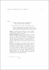
- Purpose : To investigate ultrastructural changes of the mouse lung induced by whole lung gamma irradiation and to evaluate the effect of prophylactic administration of steroid against acute lung injury. Materials and Methods :. One hundred and twenty ICR mice were used and whole lung was irradiated with telecobalt machine. Whole lung doses were 8 and 12Gy, and 10mg of methyl prednisolone was administrated intraperitoneally for two and four weeks. At the end of the observation period, mice were sacrificed by cervical dislocation. The lungs were removed and fixed inflated. Histopathological examination of acute radiation injuries were Performed by light microscopic and transmission electron microscopic examination. Results : Control group with BGy is characterized by damage to the type I Pneumocyte and the endothelial cell of the capillary. edema of alveolar wall and interstitium. and fibroblast proliferation. Control group with 120y is characterized by more severe degree of type 1 pneumocyte damage and more prominant inflammatory cell infiltration. Destructed cell debris within the alveolar space were also noted After steroid administration, 8Gy experimental group showed decreased degree of inflammatory reactions but fibroblast proliferation and basal lamina damages were unchanged. Experimental group with 12Gy showed lesser degree of inflammatory reactions similar to changes of 8Gy experimental group. Conclusion : These studies suggest that the degree of interstitial edema and inflammatory changes were related to radiation dose but Proliferation of the fibroblast and structural changes of basal lamina were not related to radialion dose. Experimental administration of steroid for 2 to 4 weeks after whole lung irradiation suggest that steroid can suppress alveolar and endothelial damages induced by whole lung irradiation but Proliferation of the fibroblast and structural changes of basal lamina were not related to administration of steroid.
목적 : 방사선량에 EK른 급성폐손상을 병리조직학적으로 분석하고 예방목적으로 투요한 스테로이드의 효과를 확인하여 방사선에 의한 폐손상의 감소방안을 모색하고자 한다. 대상 및 방법 : 생후 30일 정도인 체중 25+2g의 ICR 마우스 120마리를 암수 구별없이 사용하였고, 대조군은 전폐선량이 각각 8Gy와 12Gy인 방사선조사만 시행한 군과 방사선조사와 생리식염수를 투여한 또다른 군으로 하엿으며, 실험군은 방사선조사와 스테로이드를 복강내로 투여한 군으로 하여 방사선량과 스테로이드투여에 따른 급성폐손상의 양상을 광학 현미경 및 투과 전자현미경을 이용하여 병리조직학적으로 분석하였다. 결과 : 8Gy 대조군에서는 경미한 염증세포의 침윤, I형 폐포상피세포는 세포질이 파괴되거나 폐포강내로 돌출하였으며, 폐포 모세혈관은 경미하게 확장되었고 내피세포의 세포질과 간질은 경한 부종을 보였다. 8Gy 실험군에서 대조군에 보였던 폐포상피세포 및 내피 세포의 변화가 정도가 매우 약하였으나 섬유모세포의 증식과 기저판의 파괴정도는 대조군과 비슷하였다. 12Gy 대조군에서는 8Gy 대조군과 비교하여 염증세포의 침윤이 증가되었고 폐포강내에는 파기된 조직 파편이 산재해 있었으며 폐실질의 출혈과 무기폐가 관찰되었다. 12Gy 실험군에서는 대조군에서 보이던 현저한 염증변화가 많이 감소하였으며 폐포벽의 부종과 무기폐는 대조군과 비교하여 많이 감소되었으나 섬유모세포의 증식과 기저판의 왜곡 및 파괴는 차이가 없었다. 결론 : 방사선조사에 의한 급성폐Ths상은 전폐조사량이 8Gy에서 12Gy로 증가함에 따라 염증 세포의 침윤, 폐포상피세포의 손상, 모세혈관 내피세포의 손상, 간질의 부종정도가 증가하였으며, 방사선조사후 스테로이드를 2주일에서 4주일간 투여한 결과 이들 소견이 많이 감소하였으나 섬유모세포의 증식정도와 기저판의 변화는 조사된 방사선량이나 스테로이드 투여여부와 무관하였다.
- Alternative Title
- The Effects of Steroid on Acute Lung Injury in the Mouse Induced by Whole Lung Irradiation
- Keimyung Author(s)(Kor)
- 권건영
- Publisher
- School of Medicine
- Citation
- 성낙관 et al. (1997). 전폐조사로 유발된 마우스의 급성폐손상에 대한 스테로이드의 효과. 대한치료방사선과학회지, 15(1), 37–46.
- Type
- Article
- ISSN
- 1225-6765
- Appears in Collections:
- 1. School of Medicine (의과대학) > Dept. of Pathology (병리학)
- 파일 목록
-
-
Download
 oak-bbb-3842.pdf
기타 데이터 / 471 kB / Adobe PDF
-
Items in Repository are protected by copyright, with all rights reserved, unless otherwise indicated.