KUMEL Repository
1. Journal Papers (연구논문)
1. School of Medicine (의과대학)
Dept. of Nuclear Medicine (핵의학)
유방암 환자의 전초림프절 생검에서 유방림프신티그라피와 수술 중 감마프로우브의 유용성
- Keimyung Author(s)
- Zeon, Seok Kil; Kim, You Sah
- Journal Title
- 대한핵의학회지
- Issued Date
- 2000
- Volume
- 34
- Issue
- 6
- Keyword
- Tc-99m antimony sulfide colloid; Lymphoscintigraphy; Intraoperative radioguided gamma probe; Sentinel lymph node; Breast cancer
- Abstract
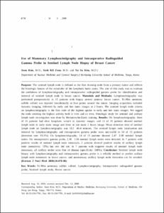
- Purpose: The sentinel lymph node is defined as the first draining node from a primary tumor and reflects the histologic feature of the remainder of the lymphatic basin status. The aim of this study was to evaluate the usefulness of lymphoscintigraphy and intraoperative radioguided gamma probe for identification and removal of sentinel lymph node in breast cancer. Materials and Methods: Lymphoscintigraphy was performed preoperatively in 15 patients with biopsy proven primary breast cancer. Tc-99m antimony sulfide colloid was injected intradermally at four points around the tumor. Imaging acquisition included dynamic imaging, followed by early and late static images at 2 hours. The sentinel lymph node criteria on lymphoscintigraphy is the first node of the highest uptake in early and late static images. We tagged the node emitting the highest activity both in vivo and ex vivo. Histologic study for sentinel and axillary lymph node investigation was done by Hematoxylin-Eosin staining. Results: On lymphoscintigraphy, three of 15 patients had clear lymphatic vessels in dynamic images, and 11 of 15 patients showed sentinel lymph node in early static image and three in late static 2 hours image. Mean detection time of sentinel lymph node on lymphoscintigraphy was 33.5¡¾48.4 minutes. The sentinel lymph node localization and removal by lymphoscintigraphy and intraoperative gamma probe were successful in 14 of 15 patients (detection rate: 93.3%). On lymphoscintigraphy, 14 of 15 patients showed 2.47¡¾2.00 sentinel lymph nodes. On intraoperative gamma probe, 2.36¡¾1.96 sentinel lymph nodes were detected. In 7 patients with positive results of sentinel lymph node metastasis, 5 patients showed positive results of axillary lymph node (sensitivity: 72%) but two did not. In 7 patients with negative results of sentinel lymph node metastasis, all axillary nodes were free of disease (specificity: 100%). Conclusion: Sentinel lymph node biopsy with lymphoscintigraphy and intraoperative gamma probe is a reliable method to predict axillary lymph node metastasis in breast cancer, and unnecessary axillary lymph node dissection can be avoided.
목적: 조직 검사에서 유방암으로 확진된 환자 15명
(평균 연령 50.4세)을 대상으로 수술 전에 시행한
유방림프신티그라피(lymphoscintigraphy)와 수술 중
감마프로우브를 이용하여, 림프관 유입형태 및 전
초림프절(sentinel lymph node)을 찾아, 전초 및 액
와림프절을 각각 절제, 생검하여, 전초 림프절의 림
프신티그라피 발현율, 전초림프절 전이와 액와림프
절 전이의 상관 관계 등을 보고자 하였다. 대상 및
방법: 환자의 임상병기는 병기 I-II 이었고, 4례에
서 액와림프절이 촉지되었다. 침습성 관암 13명, 수
질암 및 포도당 풍부암이 각각 1명씩이었다. 유방
림프신티그라피는 다음과 같이 시행하였다. 방사성
의약품 Tc-99m antimony sulfide colloid 30∼37
MBq을 총 0.4 ml 용량으로 만들어, 원발 종괴에서
2∼3 mm 떨어진 위치의 12, 3, 6, 9시 방향에 각각
0.1 ml를 피내 주사하고 약 2분 동안 마사지하였다.
저에너지, 고해상도 평행 조준기를 이용하여 초기
동적 영상(10 sec/frame)을 10분간 시행하였으며,
이어서 5분 간격으로 30∼60분에 걸쳐 초기 정적영
상을 얻었고, 주사 후 2시간에 지연영상을 획득하
였으며, 각각의 영상을 비교하여 전초림프절과 유
입 림프관을 확인하였다. 유방림프신티그라피검사
가 끝나면 즉시 수술실로 옮겨 전초림프절이라고
판독된 부위를 감마프로우브로 찾아 림프절의 계수
와 배후 방사능을 측정하였고, 이 부위를 절개하여
조직을 떼어내 표지하고 생검하였으며, 그 외에 배
후 방사능보다 높은 계수를 보인 부위가 있으면 따
로 표지하여 조직 검사를 하였다. 모든 환자에서 원
발 종양의 절제술과 액와림프절 절제술을 시행하였
다. 결과: 전체 환자 15명 가운데 14명에서 유방림
프신티그라피 및 수술 중 감마 프로우브로 전초림
프절이 발견되었다(전초 림프절 검출율: 93.3%). 유
방림프신티그라피로 발견된 평균 전초림프절수는
2.47±2.00개였으며, 감마프로우브를 이용하여 수
술로 절제된 평균 전초림프절 수는 2.36±1.96개였
다. 초기 동적 유방림프신티그라피에서 전초림프절
로 유입되는 림프관이 총 15명 중 3명에서 관찰 할
수 있었으며(20%), 3명에서는 전초림프절이 2시간
지연 영상에서만 발견되었다(20%). 유방림프신티그
라피에서 전초림프절이 나타난 시간은 평균 33.4±
48.4분이었다. 전초림프절의 조직 생검 결과 14명
가운데 7명의 전초림프절에서 전이 소견이 관찰되
었고(50%), 이 중 5명 환자의 액와림프절에서 전이
가 보였다(예민도: 71.2%). 그러나 전초림프절에 전
이가 있었던 7명 가운데 2명은 액와림프절에서 전
이소견은 관찰되지 않았다. 전초림프절에 전이가
없었던 7명 환자에서는 모두 액와림프절에서도 전
이 소견을 관찰 할 수 없었다(특이도: 100%). 유방
림프신티그라피 및 수술 중 감마프로우브로 전초림
프절을 발견 할 수 없었던 1명에서 액와절제술 후
액와림프절 조직에서 림프절에 전이가 관찰되었다.
결론: 따라서 유방암 환자에서 유방림프신티그라피
와 수술 중 감마프로우브를 이용한 전초림프절 생
검은 액와림프절 전이 평가에 있어 높은 예민도와
특이도를 나타내므로 불필요한 액와림프절 절제술
을 줄이는데 도움이 될 것이다.
- Alternative Title
- Use of Mammary Lymphoscintigraphy and Intraoperative Radioguided Gamma Probe in Sentinel Lymph Node Biopsy of Breast Cancer
- Publisher
- School of Medicine
- Citation
- 김순 et al. (2000). 유방암 환자의 전초림프절 생검에서 유방림프신티그라피와 수술 중 감마프로우브의 유용성. 대한핵의학회지, 34(6), 478–486.
- Type
- Article
- ISSN
- 1225-6714
- Appears in Collections:
- 1. School of Medicine (의과대학) > Dept. of Nuclear Medicine (핵의학)
1. School of Medicine (의과대학) > Dept. of Surgery (외과학)
- 파일 목록
-
-
Download
 oak-bbb-4139.pdf
기타 데이터 / 155.46 kB / Adobe PDF
-
Items in Repository are protected by copyright, with all rights reserved, unless otherwise indicated.