KUMEL Repository
1. Journal Papers (연구논문)
1. School of Medicine (의과대학)
Dept. of Anesthesiology & Pain Medicine (마취통증의학)
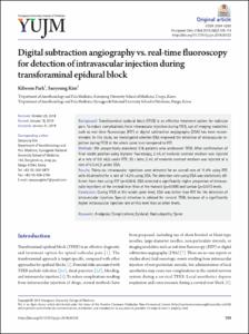
Digital subtraction angiography vs. real-time fluoroscopy for detection of intravascular injection during transforaminal epidural block
- Keimyung Author(s)
- Park, Ki Bum
- Journal Title
- Yeungnam University Journal of Medicine
- Issued Date
- 2019
- Volume
- 36
- Issue
- 2
- Keyword
- Analgesia; Complications; Epidural; Radiculopathy; Spine
- Abstract
- Background:
Transforaminal epidural block (TFEB) is an effective treatment option for radicular pain. To reduce complications from intravascular injection during TFEB, use of imaging modalities such as real-time fluoroscopy (RTF) or digital subtraction angiography (DSA) has been recommended. In this study, we investigated whether DSA improved the detection of intravascular injection during TFEB at the whole spine level compared to RTF.
Methods:
We prospectively examined 316 patients who underwent TFEB. After confirmation of final needle position using biplanar fluoroscopy, 2 mL of nonionic contrast medium was injected at a rate of 0.5 mL/s under RTF; 30 s later, 2 mL of nonionic contrast medium was injected at a rate of 0.5 mL/s under DSA.
Results:
Thirty-six intravascular injections were detected for an overall rate of 11.4% using RTF, with 45 detected for a rate of 14.2% using DSA. The detection rate using DSA was statistically different from that using RTF (p=0.004). DSA detected a significantly higher proportion of intravascular injections at the cervical level than at the thoracic (p=0.009) and lumbar (p=0.011) levels.
Conclusion:
During TFEB at the whole spine level, DSA was better than RTF for the detection of intravascular injection. Special attention is advised for cervical TFEB, because of a significantly higher intravascular injection rate at this level than at other levels.
- Keimyung Author(s)(Kor)
- 박기범
- Publisher
- School of Medicine (의과대학)
- Citation
- Kibeom Park and Saeyoung Kim. (2019). Digital subtraction angiography vs. real-time fluoroscopy for detection of intravascular injection during transforaminal epidural block. Yeungnam University Journal of Medicine, 36(2), 109–114. doi: 10.12701/yujm.2019.00122
- Type
- Article
- ISSN
- 2384-0293
- Source
- https://yujm.yu.ac.kr/journal/view.php?number=2422
- Appears in Collections:
- 1. School of Medicine (의과대학) > Dept. of Anesthesiology & Pain Medicine (마취통증의학)
- 파일 목록
-
-
Download
 oak-2019-0257.pdf
기타 데이터 / 635.17 kB / Adobe PDF
-
Items in Repository are protected by copyright, with all rights reserved, unless otherwise indicated.